A breath-hold R2 mapping pulse sequence detects a decrease in myocardial ferritin iron after one-week of iron chelation
- PDF / 809,399 Bytes
- 2 Pages / 610 x 792 pts Page_size
- 69 Downloads / 260 Views
BioMed Central
Open Access
Oral presentation
A breath-hold R2 mapping pulse sequence detects a decrease in myocardial ferritin iron after one-week of iron chelation Daniel Kim*1, Ed X Wu2, Jens Jensen1, Wing-Yan Au2, Li Feng1, Jerry S Cheung2, Shau-Yin Ha2, Sujit S Sheth3 and Gary M Brittenham3 Address: 1New York University School of Medicine, New York, NY, USA, 2The University of Hong Kong, Hong Kong, Hong Kong and 3Columbia University College of Physicians and Surgeons, New York, NY, USA * Corresponding author
from 13th Annual SCMR Scientific Sessions Phoenix, AZ, USA. 21-24 January 2010 Published: 21 January 2010 Journal of Cardiovascular Magnetic Resonance 2010, 12(Suppl 1):O69
doi:10.1186/1532-429X-12-S1-O69
Abstracts of the 13th Annual SCMR Scientific Sessions - 2010
Meeting abstracts - A single PDF containing all abstracts in this Supplement is available here. http://www.biomedcentral.com/content/files/pdf/1532-429X-11-S1-infoThis abstract is available from: http://jcmr-online.com/content/12/S1/O69 © 2010 Kim et al; licensee BioMed Central Ltd.
Introduction In transfusional iron overload, almost all the excess iron is sequestered intracellularly as ferritin iron, a dispersed, soluble and rapidly mobilizable fraction, and hemosiderin iron, an aggregated, insoluble fraction that is a long-term reserve. The effective transverse relaxation rate (R2*) of myocardium is predominantly influenced by hemosiderin iron and, even with intensive iron-chelating therapy, changes only slowly over several months [1]. Intracellular ferritin iron is evidently in equilibrium with the low molecular weight cytosolic iron pool [2] that can decrease rapidly with iron chelation. We propose to use a new breath-hold fast spin-echo (FSE) [3] pulse sequence that permits calculation of RR2 [4], a "reduced transverse relaxation rate" as a measure of myocardial ferritin iron that is largely independent of hemosiderin iron.
at mid-diastole, initially after discontinuing iron-chelation for one week, and subsequently after resuming their usual therapy (group 1: deferasirox; group 2: deferoxamine and/or deferiprone), for one week. Three different sets of FSE data were acquired in separate breath-holds with different echo spacings (ESP). For details on the pulse sequence and its parameters, please see references [3,5]. A
Purpose To use RR2 measurements to detect short-term changes in myocardial ferritin iron produced by iron-chelating therapy.
Methods We imaged 10 patients with thalassemia major (New York; mean age = 26.9 ± 10.3 years) on a 1.5 T MR scanner (Siemens-Avanto), and another 8 patients with thalassemia (Hong Kong; mean age = 29.3 ± 8.6 years) on a 3 T scanner (Phillips-Achieva). Both sets of patients were imaged in a mid-ventricular short-axis plane of the heart
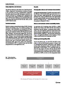
Figure discontinuing ing (Leftchelation column) 1 for chelation R2*one andweek (right for one column) week;RR (bottom (top resumrow) 2 maps:row) (Left column) R2* and (right column) RR2 maps: (top row) discontinuing chelation for one week; (bottom row) r
Data Loading...