Accuracy of freehand pedicle screws versus lateral mass screws in the subaxial cervical spine
- PDF / 1,322,473 Bytes
- 10 Pages / 595.276 x 790.866 pts Page_size
- 118 Downloads / 345 Views
CASE SERIES
Accuracy of freehand pedicle screws versus lateral mass screws in the subaxial cervical spine Hwee Weng Dennis Hey1,2 · Wen‑Hai Zhuo2 · Yong Hao Joel Tan1 · Jiong Hao Tan1 Received: 2 January 2020 / Accepted: 6 April 2020 © Scoliosis Research Society 2020
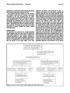
Abstract Study design Radiographic comparative study with prospectively collected data. Objectives To assess the accuracy of subaxial cervical pedicle screw (CPS) placement with freehand technique compared to lateral mass screws (LMS). Summary of background data The freehand cervical pedicle screw insertion technique guided by intraoperative lateral C-arm imaging has been shown to be both safe and effective. However, no study has performed a 100% audit of this technique using pre- and postoperative computed tomography (CT) to determine its true accuracy, as well as its reduction capability of CPS and LMS instrumentation. Methods 36 consecutive patients treated surgically by a single surgeon with the exclusive practice of LMS and subsequently CPS over 2 years were included. CT and EOS slot scanner were performed pre- and post-operatively to determine the extent of pedicle screw breach and to assess sagittal alignment reduction between CPS and LMS groups. Predictors of pedicle screw breaches were also identified using multivariate analysis. Results CPS fixation was more effective in restoring global cervical angle and had superior reduction capability of cervical lordosis at the levels of C3/4 (5.00 ± 3.92, p = 0.008), C4/5 (6.63 ± 5.5, p = 0.010) and C5/6 (7.22 ± 6.19, p = 0.004) compared to LMS fixation. Pedicle screw breaches occurred most commonly at C4 (p = 0.003), and most commonly involved the lateral pedicle wall (p 10° [6], (2) central canal stenosis > 80% [7], and (3) K-line minus in the context of ossification of the posterior longitudinal ligament [8]. All patients in the first year underwent LMS instrumentation, and all patients in the second year underwent CPS instrumentation.
Surgical methods All procedures were performed in the prone position with a Mayfield clamp and the head in a neutral position. A C-arm was positioned to allow assessment of the cervical spine using the lateral view during screw insertion. A midline approach with subperiosteal dissection was utilized for exposure. Pilot holes were made following which the intended screw track was created using a straight probe. For pedicle screw insertion, the similar technique to that described by Abumi et al. [9] was used. The entry point was located at the posterior surface of lateral mass close to the inferior edge of the cranially adjacent facet joint, and 2 mm medial to the lateral mass notch. The trajectory was between 25° and 45° of medial angulation in the axial plane. Further guidance was obtained from preoperative axial CT scan images of each vertebra to refine the medialization angle for each pedicle screw. During probing, this angle was exaggerated by 5°–10° to allow “walking off the medial wall of the pedicle”. In the sagittal plane, the direction of the pedicle
Data Loading...