Acute new-onset symptomatic seizures in the context of mild COVID-19 infection
- PDF / 589,780 Bytes
- 3 Pages / 595.276 x 790.866 pts Page_size
- 100 Downloads / 252 Views
LETTER TO THE EDITORS
Acute new‑onset symptomatic seizures in the context of mild COVID‑19 infection Maria Gaughan1 · Sean Connolly2 · Shamindra Direkze3 · Justin A. Kinsella1 Received: 28 July 2020 / Revised: 1 September 2020 / Accepted: 2 September 2020 © Springer-Verlag GmbH Germany, part of Springer Nature 2020
Dear Sirs, In the midst of the COVID-19 global pandemic, two patients with no previous history of seizures presented to our institution with acute symptomatic seizures in the context of mild respiratory SARS-CoV-2 infection.
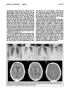
Case 1 An 87-year-old male presented to the Emergency Department (ED) at our hospital following a generalised tonic–clonic seizure, with another 30-s convulsion witnessed in ED. During a recent admission at another hospital for treatment of cellulitis, hoarseness prompted COVID-19 testing. RT-PCR amplification of SARS-Cov-2 nucleic acid from a nasopharyngeal swab was positive. He had remained otherwise asymptomatic and tested negative for SARS-CoV-2 prior to discharge. He had a history of traumatic brain injury, 32 years previously, from which he made a full recovery. Examination in ED revealed drowsiness and disorientation with no lateralising clinical signs. Urgent brain CT revealed encephalomalacia involving the inferior frontal and bilateral anterior temporal lobes consistent with previous trauma. CXR was normal. Laboratory investigations are outlined in Table 1. RT-PCR amplification of SARS-Cov-2 nucleic acid from a nasopharyngeal swab * Maria Gaughan [email protected] 1
Department of Neurology, University College Dublin, St. Vincent’s University Hospital, Elm Park, Dublin 4, Ireland
2
Department of Clinical Neurophysiology, St. Vincent’s University Hospital and University College Dublin, Dublin, Ireland
3
Department of Gastroenterology, St. Vincent’s University Hospital, Elm Park, Dublin 4, Ireland
was positive. CSF was not performed due to rapid clinical recovery. The patient remained clinically well with no respiratory symptoms throughout admission. EEG was performed following discharge. This demonstrated intermittent fronto-temporal dysfunction maximal on the right side, compatible with the known imaging abnormalities.
Case 2 A 77-year-old female presented to ED following a prolonged first generalized tonic–clonic seizure. A further generalised convulsion in the ambulance was terminated with 4 mg intravenous (IV) Lorazepam after 3 min. Collateral history revealed malaise for two days prior to presentation, but no respiratory symptoms. She had no relevant past medical history. She was high functioning at baseline, working in higher education as a course director. Urgent noncontrast CT brain and CT angiography was normal. CXR demonstrated bibasal infiltrates. RT-PCR amplification of SARS-Cov-2 nucleic acid from a nasopharyngeal swab was positive. CSF constituents were normal (Table 1) and SARS-CoV-2 in CSF was not detected. MRI Brain was normal. The patient had a prolonged encephalopathy with disorientation and bradyphrenia resulting in a protracted ad
Data Loading...