Anatomical analysis of mandibular posterior teeth for endodontic microsurgery: a cone-beam computed tomographic evaluati
- PDF / 1,516,552 Bytes
- 7 Pages / 595.276 x 790.866 pts Page_size
- 19 Downloads / 369 Views
ORIGINAL ARTICLE
Anatomical analysis of mandibular posterior teeth for endodontic microsurgery: a cone-beam computed tomographic evaluation Kug Jin Jeon 1 & Chena Lee 1 & Yoon Joo Choi 1 & Sang-Sun Han 1 Received: 21 May 2020 / Accepted: 1 September 2020 # Springer-Verlag GmbH Germany, part of Springer Nature 2020
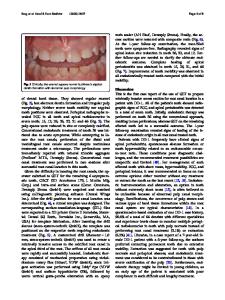
Abstract Objective The purpose of this study was to analyze the anatomical structures relevant for endodontic microsurgery in the mandibular posterior teeth using a cone-beam computed tomography (CBCT). Material and methods A total of 963 mandibular posterior teeth were analyzed in CBCT scans from 133 patients. The buccolingual and mesiodistal dimensions of the root and the buccal bone thickness overlying the root were measured at the site of root resection (apical 3 mm). At this location, the relationship between the buccal cortical bone and root was classified into three types (separated, contact, and exposed), and the distance from the root apex to the mandibular canal was measured. Results The thickest buccolingual dimension of the roots was found in the mesial roots of first molars, at 5.59 ± 0.97 mm. The buccal bone thickness overlying the root became thicker in posterior tooth locations. In the first premolar and first molar mesial root, contact was the most common type of relationship between the buccal cortical bone and root. As the position of the teeth became more posterior, the distance from the apex to the mandibular canal became shorter. Conclusions As the position of the teeth became more posterior, the buccal bone thickness increased and the distance to the mandibular canal became closer; therefore, particular attention is required for posterior teeth. The first premolar and the first molar mesial root are often in contact with the buccal cortical bone, which may allow infections to spread to the buccal structure more easily and negatively affect for post-surgical healing. Clinical relevance When planning and performing endodontic microsurgery, understanding the anatomical structure of the surgical site will help minimize tissue damage and reduce complications. Keywords Cone-beam computed tomography . Endodontics . Mandible . Microsurgery . Tooth
Introduction Due to advances in implant technology, it has become increasingly common for implants to be placed after tooth extraction; nonetheless, maintenance of the natural teeth is the dentist’s ultimate goal. To preserve the natural teeth, endodontic surgery may be an option when conventional endodontic treatment fails or endodontic retreatment is inappropriate. Mandibular posterior teeth are difficult to access with
Kug Jin Jeon and Chena Lee contributed equally to this work. * Sang-Sun Han [email protected] 1
Department of Oral and Maxillofacial Radiology, Yonsei University College of Dentistry, 50-1 Yonsei-ro Seodaemun-gu, Seoul 03722, Korea
instruments, their buccal cortical bone is thick, and they are close to the inferior alveolar canal; collectively, these factors result in a lower success rate and an increased risk of complications
Data Loading...