Angioscopic findings of organized thrombosis of iliac vein in patient with chronic thromboembolic pulmonary hypertension
- PDF / 904,532 Bytes
- 2 Pages / 595.276 x 790.866 pts Page_size
- 110 Downloads / 327 Views
IMAGES IN CARDIOVASCULAR INTERVENTION
Angioscopic findings of organized thrombosis of iliac vein in patient with chronic thromboembolic pulmonary hypertension Naoki Kubota1 · Kazuyuki Ozaki1 · Takahiro Hakamata1 · Yasuhiro Ikami1 · Makoto Hoyano1 · Tohru Minamino1 Received: 30 August 2020 / Accepted: 15 October 2020 © Japanese Association of Cardiovascular Intervention and Therapeutics 2020
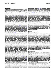
Keywords Angioscopy · Deep-vein thrombosis · Organized thrombus · Chronic thromboembolic pulmonary hypertension A 55-year-old woman with chronic thromboembolic pulmonary hypertension (CTEPH) was admitted to our hospital for balloon pulmonary angioplasty (BPA). She had a history of deep-vein thrombosis (DVT) in left lower limb 13 years ago and treated with warfarin. Contrast-enhanced computed tomography (CT) showed narrowing of left common iliac vein (CIV) (Fig. 1a). Venography showed slit-like contrast defect in the left CIV and external iliac vein (EIV) (Fig. 1b). Angioscopy (Forwardlooking®, OVALIS, Osaka, Japan) was performed during BPA. A 6-Fr guiding catheter was inserted from right common femoral vein and advanced to left EIV. Angioscopy revealed “slit” and “mesh” appearance in the left EIV (Fig. 1c, d).
CTEPH is considered to occur following history of acute pulmonary embolism and DVT. In Japanese registry [1], however, only almost half of patients with CTEPH had DVT episode and the relationship between CTEPH and DVT is still uncertain. Organized thrombus (“mesh,” and “slit” appearance) is often observed on angioscopy of pulmonary arteries in the setting of chronic thromboembolic pulmonary hypertension (CTEPH) [2]. In our case, the organized thrombus in veins was similar to that in pulmonary arteries in the setting of CTEPH. Our findings were interesting that organized thrombus were also seen in veins with CTEPH patient.
Electronic supplementary material The online version of this article (https://doi.org/10.1007/s12928-020-00724-9) contains supplementary material, which is available to authorized users. * Naoki Kubota [email protected]‑u.ac.jp 1
Department of Cardiovascular Biology and Medicine, Niigata University Graduate School of Medical and Dental Sciences, 1‑757, Asahimachidori, Chuo‑ku, Niigata 951‑8510, Japan
13
Vol.:(0123456789)
N. Kubota et al.
Fig. 1 a Computed tomography at admission showed narrowing of left common iliac vein (CIV) (arrow). b Venography showed slit-like contrast defect in the left CIV and external iliac vein (EIV). c Angi-
oscopy findings at red arrow in b. d Angioscopy findings at yellow arrow in b
Compliance with ethical standards
References
Conflict of interest The authors have no conflicts of interest to declare.
1. Tanabe N, Sugiura T, Tatsumi K. Recent progress in the diagnosis and management of chronic thromboembolic pulmonary hypertension. Respir Investig. 2013;51:134–46. 2. Takano T, Ozaki K, Hoyano M, Yanagawa T, Kashimura T, Minamino T. Angioscopic findings during balloon pulmonary angioplasty in chronic thromboembolic pulmonary hypertension. Card
Data Loading...