Applying electrical impedance tomography to dynamically monitor retroperitoneal bleeding in a renal trauma patient
- PDF / 296,416 Bytes
- 2 Pages / 595.276 x 790.866 pts Page_size
- 60 Downloads / 280 Views
CO RRESPONDENCE
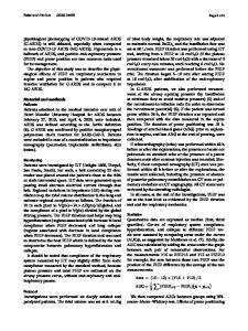
tool to continuously monitor patients and identify ARB. We present the first case report of the use of electrical impedance tomography (EIT), a noninvasive, dynamic and functional imaging modality, in the monitoring of ARB under clinical conditions. A 27-year-old man was diagnosed with grade IV injury to the left kidney by CT and B-scan ultrasonography, and was put on close clinical observation as conservative treatment. As Applying electrical impedance well as conventional monitoring of vital signs, including blood pressure tomography to dynamically (BP), heart rate (HR) and SpO2, the monitor retroperitoneal patient was also continuously monitored by EIT for 9 h. bleeding in a renal trauma The patient was kept lying supine patient quietly and 16 disposable Ag–AgCl electrodes were attached around the Accepted: 28 February 2013 body 2 cm below the injury and Published online: 29 March 2013 Ó The Author(s) 2013. This article is covering the area of the kidney. The published with open access at electrodes were connected to an Springerlink.com elastic belt to ensure good contact with the skin. The area of the injury, the electrodes and the elastic belt are shown in Fig. 1. The EIT data acquisition system was developed by Dear Editor, the FMMU EIT Group [3–5]. A Active retroperitoneal bleeding stimulus frequency of 50 kHz, a (ARB) is characterized by a large amount of bleeding over a long period constant driving current of 1 mA, the polar driving and adjacent measureof time. Massive hemorrhage from ment mode, and an imaging speed of ARB, if not detected early enough, one frame per second were applied. may lead to serious consequences including kidney resection and even An optimized EIT image reconstrucdeath [1, 2]. But up to now there has tion strategy was applied using a damped least-squares method to been no effective clinical imaging Fusheng You Xuetao Shi Wanjun Shuai Hongyi Zhang Wei Zhang Feng Fu Ruigang Liu Canhua Xu Tingyi Bao Xiuzhen Dong
Fig. 1 Photographs showing the area of the injury, the EIT electrodes and the belt
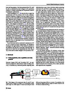
improve the spatial resolution of the reconstructed images [5]. The reconstructed dynamic EIT images were integrated with one of the patient’s CT images which acted as the static background image. The results of the urine test confirmed gross hematuria and renal injury. The results of the blood tests were within normal ranges except that the WBC was high. There were no significant changes in vital signs (BP, HR, and SpO2). The fused EIT/CT images during the 9 h of EIT monitoring are shown in Fig. 2. At about 1330 hours the local image in the left renal region began to become red because of a decrease in impedance caused by the ARB. Up to about 1730 hours, the region showing the impedance decrease was almost fixed over the left renal area, and gradually deepened in color and increased in size. As the EIT monitoring showed a continuous decrease in resistivity and ARB, a CT scan was performed, immediately followed by digital subtraction angiography (DSA) of the celiac artery and th
Data Loading...