Asymmetric Lattice Response During Tensile and Compressive Deformation of a Uranium-Niobium Shape Memory Alloy
- PDF / 639,910 Bytes
- 6 Pages / 612 x 792 pts (letter) Page_size
- 75 Downloads / 394 Views
Q7.6.1
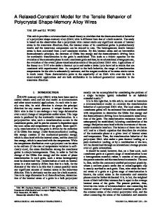
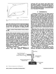
Asymmetric Lattice Response During Tensile and Compressive Deformation of a UraniumNiobium Shape Memory Alloy D. W. Brown, M. A. M Bourke, P. S. Dunn, R. D. Field, M. G. Stout, D. F. Teter, D. J. Thoma, S. C. Vogel Los Alamos National Laboratory, Los Alamos, NM, 87545 ABSTRACT The deformation of polycrystalline uranium 6 wt. % niobium (U6Nb) was studied in-situ during uniaxial tensile and compressive deformation by time-of-flight neutron diffraction. Diffraction patterns were recorded at incremental strains to roughly 4% total deformation. The asymmetry in the crystallographic response of the lattice is discussed. Introduction Depending on the Nb concentration, U-Nb alloys exhibit the following room temperature structures on quenching from the γ phase; α′ orthorhombic (0-10 at.%, 0-4.2 wt.%); α′′ monoclinic (10-16 at. %, 4.2-6.9 wt.%): γº tetragonal (16-20 at. %, 6.9-8.9 wt.%) [1-5], using the notation suggested by Lehmann et al. [6]. The uranium-rich U-Nb alloys in the α′′ and γ0 phases exhibit the shape memory effect (SME), i.e., the ability to recover deformation-induced strains by subsequent heating. Jackson et al. first reported SME in a U-Nb alloy in 1978 [7]. A systematic phenomenological study of the SME as a function of Nb concentration between 5.8 and 7.6 wt. % demonstrated the SME in both the α′′ and γº phases, although larger strain recovery was observed in the α′′ phase [8]. The shape memory effect results from the preferred growth of one or more martensite variants with transformation strains that can accommodate the imposed strain through twin boundary motion. A previously published neutron diffraction study [9] focused on in-situ determination of lattice strain and development of the twinned microstructure of martensitic U6Nb in tension. The present study extends the past study to include compressive deformation and examines the asymmetric response of the crystallographic lattice of the material in tension and compression. Experimental Details The details of the sample preparation and microstructure are presented in earlier publications [9, 10]. In-situ neutron diffraction measurements were performed on the Neutron Powder Diffractometer (NPD) and SMARTS diffractometer at the Manuel Lujan Jr. Neutron Scattering Center, LANSCE, Los Alamos National Laboratory. The NPD and SMARTS are very similar in terms of diffraction geometry, e.g. incident and scattered flight path, and details are published elsewhere [11, 12]. In both diffractometers, a pulsed neutron beam with a continuous wavelength spectrum (0.4 - 4Ǻ) impinges on the sample and scattered in all directions. Detectors banks consisting of 3He filled tubes are located 1.5 m from the sample situated at ± 90º relative to the incident beam. Quasi-static loading is performed on a horizontal Instron® load frame with the load axis oriented at 45° relative to the incident beam. The instrument geometry combined with
Q7.6.2
the continuous energy spectrum of the incident beam provides two complete diffraction patterns collected simultaneously with d
Data Loading...