Bilateral simultaneous quadriceps tendon rupture in a patient with diabetes and gout: a case report and review of the li
- PDF / 384,946 Bytes
- 7 Pages / 595.276 x 790.866 pts Page_size
- 52 Downloads / 345 Views
CASE REPORT
Bilateral simultaneous quadriceps tendon rupture in a patient with diabetes and gout: a case report and review of the literature K. Moens & S. Harth & Y. Depaepe & A. Harth & G. Vandendriessche & C. Van Vynckt
Received: 16 July 2012 / Accepted: 27 March 2013 # EFORT 2013
Introduction A quadriceps tendon rupture (QTR) is a well-known condition in orthopedic practice, requiring adequate surgical treatment to avoid residual deformity and loss of knee function [23]. Bilateral spontaneous rupture of the quadriceps tendon is rather rare [4]. It is usually associated with concomitant underlying conditions such as advanced age, chronic renal failure, obesity, diabetes mellitus, gout, hyperparathyroidism, pseudogout, steroid usage, and autoimmune diseases such as rheumatoid arthritis and systemic lupus erythematosus [4]. Chronic renal failure is considered to be the most frequently encountered risk factor in simultaneous QTR with a reported rate of more than 40 % [23]. In Table 1, case reports of bilateral quadriceps tendon ruptures that were found in literature are listed. The commonest cause of bilateral simultaneous rupture appears to be a sudden violent contraction of the quadriceps mechanism with the knees slightly flexed and the feet fixed [20]. It usually occurs in an effort to prevent a fall or to lift something or simply in descending stairs [26]. Shah stated that falls are the main cause (76 %) and that the commonest site of rupture is the osseotendinous junction (60 %) [27]. These ruptures tend to occur in older age groups typically more than 40 years of age. In older patients the rupture occurs typically at the osteotendinous junction while in younger patients, it is more often at the mid-tendinous area [4]. Older patients had ruptured tendons from falling down, whereas younger patients had spontaneous rupture. Patients with renal or endocrine disorders, or with multiple diseases, were younger [28]. K. Moens (*) : S. Harth : Y. Depaepe : A. Harth : G. Vandendriessche : C. Van Vynckt Department of Orthopedic Surgery and Traumatology, AZ Jan Palfijn Ghent, Henri Dunantlaan 5, 9000 Ghent, Belgium e-mail: [email protected]
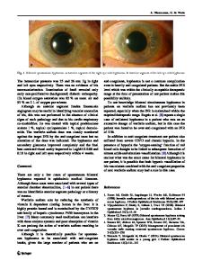
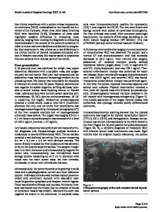
Early diagnosis is important in the management of complete quadriceps tendon rupture, because primary surgical repair is necessary within the first 48 to 72 h to preserve the extensor mechanism of the knee joint [11]. The clinical manifestation includes the triad of anterior knee pain, inability to extend the knee, and a palpable suprapatellar gap (Fig. 1) which is the so-called sulcus sign or gap test [28, 29]. Plain radiography can be useful, but are of limited diagnostic value in QTR, as it is often nonspecific or it reveals only indirect signs of the disruptions, such as a low riding or forward tilting of the patella (Fig. 2), calcifications in the substance of the tendon, a suprapatellar or joint effusion, suprapatellar calcifications due to systemic disease or due to bony transformation of scar tissues, or perhaps a poorly defined soft tissue shadow of the tendon [16, 23].
Data Loading...