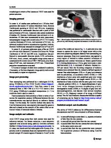
Can computed tomography-based radiomics potentially discriminate between anterior mediastinal cysts and type B1 and B2 t
- PDF / 1,517,210 Bytes
- 14 Pages / 595.276 x 790.866 pts Page_size
- 91 Downloads / 204 Views
BioMedical Engineering OnLine Open Access
RESEARCH
Can computed tomography‑based radiomics potentially discriminate between anterior mediastinal cysts and type B1 and B2 thymomas? Lulu Liu1,2,3, Fangxiao Lu1,2,3, Peipei Pang4 and Guoliang Shao1,2,3*
*Correspondence: [email protected] 3 Department of Radiology, Zhejiang Cancer Hospital, No. 1 Banshan Street, Gongshu District, Hangzhou 321022, Zhejiang, People’s Republic of China Full list of author information is available at the end of the article
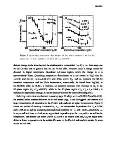
Abstract Background: Anterior mediastinal cysts (AMC) are often misdiagnosed as thymomas and undergo surgical resection, which caused unnecessary treatment and medical resource waste. The purpose of this study is to explore potential possibility of computed tomography (CT)-based radiomics for the diagnosis of AMC and type B1 and B2 thymomas. Methods: A group of 188 patients with pathologically confirmed AMC (106 cases misdiagnosed as thymomas in CT) and thymomas (82 cases) and underwent routine chest CT from January 2010 to December 2018 were retrospectively analyzed. The lesions were manually delineated using ITK-SNAP software, and radiomics features were performed using the artificial intelligence kit (AK) software. A total of 180 tumour texture features were extracted from enhanced CT and unenhanced CT, respectively. The general test, correlation analysis, and LASSO were used to features selection and then the radiomics signature (radscore) was obtained. The combined model including radscore and independent clinical factors was developed. The model performances were evaluated on discrimination, calibration curve. Results: Two radscore models were constructed from the unenhanced and enhanced phases based on the selected four and three features, respectively. The AUC, sensitivity, and specificity of the enhanced radscore model were 0.928, 89.3%, and 83.8% in the training dataset and 0.899, 84.6%, and 87.5% in the test dataset (higher than the unenhanced radscore model). The combined model of enhanced CT including radiomics features and independent clinical factors yielded an AUC, sensitivity and specificity of 0.941, 82.1%, and 94.6% in the training dataset and 0.938, 92.3%, and 87.5% in the test dataset (higher than the unenhanced combined model and enhanced radscore model). Conclusions: The study suggested the possibility that the combined model in enhanced CT provided a potential tool to facilitate the differential diagnosis of AMC and type B1 and B2 thymomas. Keywords: Anterior mediastinal cysts, Thymomas, Radiomics, Enhanced CT
© The Author(s) 2020. This article is licensed under a Creative Commons Attribution 4.0 International License, which permits use, sharing, adaptation, distribution and reproduction in any medium or format, as long as you give appropriate credit to the original author(s) and the source, provide a link to the Creative Commons licence, and indicate if changes were made. The images or other third party material in this article are included in the article’s Creative Commons licence, unles
Data Loading...