Challenges in catheter ablation of deep myocardial substrate for ventricular tachycardia
- PDF / 2,520,162 Bytes
- 3 Pages / 595.276 x 790.866 pts Page_size
- 105 Downloads / 290 Views
CASE REPORTS
Challenges in catheter ablation of deep myocardial substrate for ventricular tachycardia Anna F. Thomsen 1 & Steen Pehrson 1 & Xu Chen 1 & Christian Jons 1 & Peter Karl Jacobsen 1 Received: 14 April 2020 / Accepted: 5 June 2020 # Springer Science+Business Media, LLC, part of Springer Nature 2020
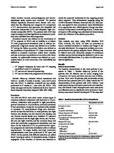
1 Case report Myocardial substrate for ventricular tachycardia (VT) may be intramural and challenging to reach with ablation in thick parts of the myocardium of the left ventricle (LV) as our case demonstrates. A 76-year-old man with ischemic heart disease, previous CABG operation, presented with recurrent episodes of sustained monomorphic VT despite amiodarone. The 12-lead ECG before initial ablation (Fig. 2A) showed ventricular tachycardia with RBBB morphology, superior axis, and cycle length 500 ms, and was suggestive of a mid-myocardial focus. Ablation was planned. A 3D high-resolution late activation time (LAT) map of the LV was performed during VT suggesting an area of deep myocardial basal and inferior/posterior located scar tissue (Fig. 1, first procedure and Online Resource, Video S1). Entrainment pacing maneuvers confirmed the findings of the LAT map. Ablation using both remote magnetic navigation (RMN) and manual ablation in the same area was acutely successful with non-inducible VT at the end. Yet, the patient developed recurrent VT with similar
Electronic supplementary material The online version of this article (https://doi.org/10.1007/s10840-020-00796-w) contains supplementary material, which is available to authorized users. * Anna F. Thomsen [email protected] 1
Department of Cardiology, Rigshospitalet, University Hospital, Copenhagen, Denmark
ECG-12 morphology (Fig. 2B) a few days after the first ablation and 3 months after the second, and was re-ablated 1 week and 3 months after first-time ablation. Third procedure was optimized to create deeper myocardial ablation lesions. (1) Retrograde endocardial access and manual catheter ablation were chosen in order to improve accessibility and contact force respectively. (2) Hypotonic external irrigation was chosen to enhance lesion formation. By decreasing ionic concentration and charge density during ablation using half-normal saline instead of normal saline, deeper myocardial lesions can be created [1]. The main benefit of using half-normal saline instead of, for example, needle ablation is that it is simple and safe. A combination of these techniques eliminated VT after < 10 s with ablation in the same area as before (mean contact force 15 g and power 40 W) as illustrated in Fig. 1, third procedure, and in the animation (Online Resource, Video S2), and the patient has now been free of VT for 6 months. For ECG and intracardiac catheter electrogram from the succesful ablationsite of VT termination during third procedure, see Online Resource, Fig. 3.
J Interv Card Electrophysiol Fig. 1 Left ventricular (LV) inferior wall three-dimensional electroanatomic map performed with the PENTARAY catheter (Biosense Webster) used with the CARTO
Data Loading...