Chiari II Malformation and Syntelencephaly in a Young Woman: Coincidence or Pathogenetic Association?
- PDF / 872,358 Bytes
- 3 Pages / 595.276 x 790.866 pts Page_size
- 81 Downloads / 317 Views
Correspondence
Chiari II Malformation and Syntelencephaly in a Young Woman: Coincidence or Pathogenetic Association? T. O. Kalayci · A. Tekes · T. A. G. M. Huisman · A. Poretti
Received: 2 October 2012 / Accepted: 8 December 2012 © Springer-Verlag Berlin Heidelberg 2012
Introduction Chiari II malformation (CII) is a mid-hindbrain malformation, which occurs in children with open spinal dysraphism, typically myelomeningocele (MMC) [1]. CII is characterized by a small posterior fossa, downward vermian herniation, pontine hypoplasia, tectal beaking, hydrocephalus, and commissural abnormalities [2]. Syntelencephaly or middle interhemispheric variant of holoprosencephaly (HPE) is a rare form of HPE. It consists of a nonseparation of the posterior frontal and parietal regions of both cerebral hemispheres [3, 4]. We report on the clinical and neuroimaging findings of a patient with CII and syntelencephaly and discuss whether it is a coincidence or a pathogenetic association. Case Report A 22-year-old woman was born with an open lumbar MMC. After surgical closure of the MMC, she developed hydrocephalus. A ventriculoperitoneal shunt was implanted. Because of cord tethering, surgical untethering was performed twice. At the last follow-up, she attended high school with an individualized integrative education program because of a mild intellectual disability. She was able to walk about 200 feet with support. Her muscle strength was 4/5 in the upper and 3/5 in the lower extremities except for
A. Poretti () · T. O. Kalayci · A. Tekes · T. A. G. M. Huisman Division of Pediatric Radiology, Russell H. Morgan Department of Radiology and Radiological Science, Johns Hopkins Hospital, 600 N Wolfe Street, Nelson B-173, Baltimore MD 21287-0842, USA e-mail: [email protected]
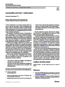
0/5 for toe dorsiflexion and hip extension/abduction. She presented with a 48-degree thoracic dextroscoliosis. Clean intermittent catheterization, oxybutynin, and a regular manual bowel emptying program were used to treat neurogenic bladder and bowel dysfunction. The last brain and spine magnetic resonance imaging (MRI) was performed at the age of 20 years on a 3 T scanner (Siemens Trio, Erlangen, Germany). Brain MRI included three-dimensional (3D) T1- and axial/coronal T2-weighted sequences and diffusion tensor imaging (DTI); spine MRI included axial and sagittal T1- and T2-weighted images (Fig. 1). MRI showed all characteristic stigmata of a CII: a small posterior fossa, mild downward herniation of the cerebellar vermis, inferior displacement of the fourth ventricle, embracement of the brainstem by the cerebellar hemispheres, small pontine prominence, mild tectal beaking, minimal medullary kinking, and prominent massa intermedia. The corpus callosum (CC) was severely abnormal: the genu and the splenium were hypoplastic, the corpus was absent. In the expected location of the callosal body, dysplastic gray and white matter was noted connecting both parts of the posterior frontal and anterior parietal lobes suggesting syntelencephaly. The transverse (right ⇔ left)
Data Loading...