Circumportal Pancreas: a Chance Encounter During Pancreaticoduodenectomy
- PDF / 1,274,439 Bytes
- 3 Pages / 595.276 x 790.866 pts Page_size
- 82 Downloads / 294 Views
CASE REPORT
Circumportal Pancreas: a Chance Encounter During Pancreaticoduodenectomy Kishore G. S. Bharathy 1 & Ankur Arora 2 & Shridhar V. Sasturkar 1 Received: 21 May 2020 / Accepted: 15 August 2020 # Association of Surgeons of India 2020
Abstract Circumportal pancreas is a rare embryological anomaly where the dorsal and ventral pancreatic anlages are fused resulting in a band of pancreatic tissue encasing the portal vein. This can be easily missed if one is not aware of the entity. This report discusses the implication of finding this anomaly during pancreaticoduodenectomy, describes the existing classification system, management strategies to complete surgery safely, and reduce the incidence of postoperative pancreatic fistula. Keywords Circumportal pancreas . Portal annular pancreas . Pancreaticoduodenectomy . Pancreatic fistula
Introduction Circumportal pancreas (CPP) is a developmental anomaly where there is abnormal fusion of the embryonic dorsal and ventral pancreatic anlages around the superior mesenteric/ portal vein [1]. It can be classified as supra-splenic, infrasplenic, or mixed type depending upon the location of the pancreatic tissue bridge in relation to the splenic vein [2]. Unless one is aware of the existence of this anomaly, it is easily missed. Presence of CPP has important implications for surgical planning and management of periampullary tumors by pancreaticoduodenectomy.
Case Report A 56-year-old lady presented with upper abdominal discomfort for 2 months. There was no jaundice, pruritus, or weight loss. She was diabetic and hypertensive, both well controlled with medications. Physical exam was unremarkable. Liver function tests showed a serum bilirubin of 1 mg/dl, AST, ALT of 82, 98 U/L respectively and alkaline phosphatase, * Shridhar V. Sasturkar [email protected] 1
Present address: Department of HPB Surgery & Liver Transplantation, Institute of Liver & Biliary Sciences, D1, Vasant Kunj, New Delhi 110070, India
2
Department of Radiology, Institute of Liver & Biliary Sciences, D1, Vasant Kunj, New Delhi 110070, India
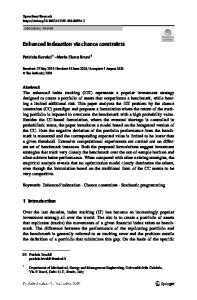
gamma-glutamyl transferase of 166 IU/L (reference range 32–92) and 262 U/L (7–64) respectively. Serum albumin was 4.3 g/dl. Ultrasound of the abdomen showed a dilated common bile duct (CBD) with intrahepatic biliary radicle dilatation. Contrast-enhanced computed tomography (CT) scan showed significant biliary dilatation but no obvious mass lesion (Fig. 1). Pancreatic duct was normal. A side viewing endoscopy followed by an endoscopic ultrasound (EUS) showed a dilated CBD with a mass measuring 1 cm at ampulla. EUS-guided fine needle aspiration confirmed adenocarcinoma. After adequate preoperative preparation, she was taken up for pancreaticoduodenectomy. At surgery after division of the pancreatic neck over the portal vein, it was found that there was a bridge of pancreatic tissue encircling the portal vein from behind and joining the pancreatic parenchyma on the left of the vessel (Fig. 2A). Careful review of the pre-operative CT scan at this point showed the pre
Data Loading...