Clinical Presentation and Therapy of Atrioventricular Septal Defect
Atrioventricular septal defects (AVSDs) consist of a number of cardiac malformations that result from abnormal development of the endocardial cushions. AVSDs occur in 0.19 of 1000 live births and constitute 4–5 % of congenital heart defects. AVSDs can be
- PDF / 263,043 Bytes
- 3 Pages / 439.37 x 666.14 pts Page_size
- 4 Downloads / 303 Views
25
David J. Driscoll
Contents 25.1 25.2 25.3 25.4 25.5 25.6
Introduction Pathologic Physiology Clinical Presentation and Physical Examination Echocardiographic and Cardiac Catheterization Issues Treatment Outcome
345 347 347 347 347 347
Abstract
Atrioventricular septal defects (AVSDs) consist of a number of cardiac malformations that result from abnormal development of the endocardial cushions. AVSDs occur in 0.19 of 1000 live births and constitute 4–5 % of congenital heart defects. AVSDs can be categorized as (1) incomplete (or partial) or (2) complete (Fig. 25.1) and (3) intermediate or transitional.
25.1
Introduction
Atrioventricular septal defects (AVSDs) consist of a number of cardiac malformations that result from abnormal development of the endocardial cushions. AVSDs occur in 0.19 of 1000 live births and constitute 4–5 % of congenital heart defects.
D.J. Driscoll Division of Pediatric Cardiology, Department of Pediatrics, Mayo Clinic College of Medicine, Rochester, MN, USA e-mail: [email protected] © Springer-Verlag Wien 2016 S. Rickert-Sperling et al. (eds.), Congenital Heart Diseases: The Broken Heart: Clinical Features, Human Genetics and Molecular Pathways, DOI 10.1007/978-3-7091-1883-2_25
345
346
a
D.J. Driscoll
b
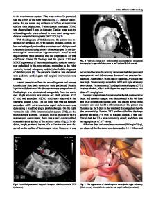
Fig. 25.1 Diagrammatic representation of a complete atrioventricular septal defect on the left. Note that instead of a mitral and a tricuspid valve, there is only one large atrioventricular valve. There are cordal attachments to the crest of the ventricular septum. The ostium primum ASD and an inlet VSD are readily apparent. On the right is an echocardiographic four-chamber view of a complete atrioventricular canal defect. The arrows represent the annulus of the common AV valve. The “X” indicates an associated ostium secundum ASD and the “*” shows the location of the ostium primum ASD. The “o” shows the location of the VSD. Abbreviations: MV mitral valve remnant, TV tricuspid valve remnant, RA right atrium, RV right ventricle, L lateral valve leaflet, A anterior leaflet, P posterior leaflet, LV left ventricle (Adapted from Feldt et al. (1976) Atrioventricular Canal Defect, WB Saunders, Philadelphia and reproduced or adapted from Driscoll, David (2006) Fundamentals of Pediatric Cardiology. Lippincott Williams & Wilkins, Baltimore with permission of the author and publisher)
AVSDs can be categorized as (1) incomplete (or partial) or (2) complete (Fig. 25.1) and (3) intermediate or transitional. Incomplete AVSDs include ostium primum ASD, common atrium, cleft mitral valve, and defects of the atrioventricular septum producing a left ventricular to right atrial shunt (Gerbode defect). Ostium primum ASD results from lack of closure of the ostium primum by the endocardial cushions. Since the endocardial cushions also form major portions of the mitral and tricuspid valves, abnormalities of the atrioventricular valves are associated with ostium primum ASDs. A cleft in the septal leaflet of the mitral valve invariably is associated with ostium primum ASDs. Complete AVSD is a con
Data Loading...