Collagen-calcium Phosphate Composite Coatings by Electrolysis-induced Self-assembly and Mineralization
- PDF / 1,256,872 Bytes
- 6 Pages / 612 x 792 pts (letter) Page_size
- 10 Downloads / 349 Views
W6.4.1
Collagen-calcium Phosphate Composite Coatings by Electrolysis-induced Self-assembly and Mineralization Yuwei Fan, Ke Duan, Rizhi Wang* Department of Metals and Materials Engineering, University of British Columbia, Vancouver, B.C. Canada V6T 1Z4 * Corresponding Author, Email: [email protected]
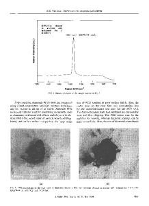
Abstract A bone-like composite coating of collagen protein and calcium phosphate minerals is considered to be bioactive and may enhance bone growth and fixation of metallic orthopedic implants. In this study, we have successfully prepared a uniform collagen fibril/octacalcium phosphate composite coating on silicon substrate by electrolytic deposition. Under a typical deposition condition, a thin (100 nm) layer of calcium phosphate coating would form on the cathode (Si) surface first, which was followed by a thick (~100 µm) composite coating. The porous composite layer consists of a collagen fibril network on which clusters of octacalium phosphate crystals nucleate and grow. The results not only provide a novel bioactive coating for biomedical implants, but also establish a new experimental protocol for studying biomineralization mechanisms of collagen based biological tissues. Introduction Bone is a natural composite mainly made from collagen fibril, carbonated apatite and small amount of non-collagen proteins. The extraordinary mechanical behavior of bone arises from its nanocomposite nature and the various levels of hierarchical structure [1]. Traditional synthetic materials for bone repair including metals, polymers and ceramics are bioinert. In searching for new generation of bone substitutes that are bioactive and biodegradable, bone-like composites made of collagen and calcium phosphate (usually hydroxyapatite) minerals have received much attention recently because they mimic the basic compositions of bone [2,3,4]. A typical method is to add base to a phosphate and calcium solution dissolved with type I collagen molecules. At a high pH (9~10), the self-assembly of collagen and the precipitation of hydroxyapatite minerals could happen simultaneously to form a nanocomposite in the solution [2, 3]. In vivo tests have proven that such bone-like composite is bioactive and biodegradable and could enhance new bone growth [2]. The drawback of the synthetic collagen/calcium phosphate composite is the low mechanical strength that limits the composite to non-loading applications. An alternative way is to apply a layer of collagen/calcium phosphate composite coating to the surface of metallic implants. One applicable example is in hip implant. Many of these artificial hip parts especially the acetabular components are fixed to bone directly (cementless hip), and a bioactive surface could accelerate
W6.4.2
bone growth and implant fixation. To apply a bone-like coating on metallic implants, a novel coating technique needs to be developed since many common coating methods such as plasma spray and sol-gel are not applicable. In this study we will introduce a new process based electrolysis deposition (ELD) to prepare
Data Loading...