Common presentation of micro hematuria and proteinuria in an uncommon disease: Questions
- PDF / 598,925 Bytes
- 2 Pages / 595.276 x 790.866 pts Page_size
- 101 Downloads / 310 Views
CLINICAL QUIZ
Common presentation of micro hematuria and proteinuria in an uncommon disease: Questions Marwa Mansour 1 & Danielle Young 1 & Mohammad Ilyas 1 & Asad Tolaymat 1 Received: 22 March 2020 / Revised: 27 March 2020 / Accepted: 31 March 2020 # IPNA 2020
Clinical presentation A 12-year-old female, previously healthy, presented to the pediatric nephrology clinic because of an incidental finding of microscopic hematuria in routine urinalysis. Repeated urinalysis showed persistent microscopic hematuria without pyuria or proteinuria. At initial presentation, the patient had no gross hematuria or dysuria. She had no swelling of the eyelids or the lower extremities. She had no hypertension. Further history revealed no bleeding tendency and no family history of kidney stones, hearing loss, or kidney disease. The patient had no arthralgia or arthritis. Initial work-up was ordered but was never done, and the patient was lost to follow-up. The patient was sent back to the nephrology clinic 8 months later due to new onset proteinuria and cola-colored urine. Urinalysis showed protein > 300 mg and significant hematuria. The rest of the urinalysis was normal. Her complete blood count showed hemoglobin 13 g/dl and normal white blood cell and platelet counts. The chemistry panel showed urea 15
The answers to these questions can be found at https://doi.org/10.1007/ s00467-020-04566-8. * Marwa Mansour [email protected] 1
University of Florida College of Medicine, Jacksonville, FL, USA
(reference range (ref) 7–20 mg/dl), creatinine 0.69 (ref 0.4– 1 mg/dl), and albumin 4 (ref 3.6–5.1 g/dl). Kidney ultrasound was normal. Urine protein-to-creatinine ratio was 1897 (ref 21– 161 mg/ g Cr) with normal calcium-to-creatinine ratio. Serum studies showed normal C3 and C4 values. Lipid profile was positive for high total cholesterol 258 (ref < 170 mg/dl), high LDL 137 (ref < 110 mg/dl), and high triglycerides 104 (ref < 90 mg/dl), HDL was normal at 100 (ref > 45 mg/dl). Autoimmune work-up, including ANCA panel screen, was normal. Lupus panel was negative. Cryoglobulins were undetectable. Infectious work-up including hepatitis B and parvovirus screen was negative; anti-streptolysin O was within normal levels. Given these results: 1. What would be your differential diagnosis? 2. What further studies would you recommend?
Pediatr Nephrol
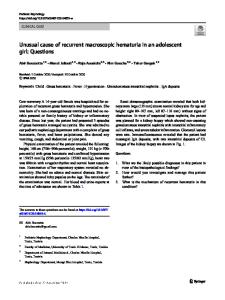
Fig. 1 Light microscopy. Diffuse endocapillary proliferation, diffuse mesangial expansion with mesangial hypercellularity. Rare duplication of the glomerular basement membranes is seen
Fig. 3 Electron microscopy. Glomerular sections show diffuse endocapillary proliferation, diffuse mesangial hypercellularity, numerous subendothelial electron-dense deposits, numerous mesangial electron-dense deposits, and rare new basement membrane formation. Moderate foot process effacement
Case continued To determine the underlying etiology, a kidney biopsy was performed that revealed membranoproliferative glomerulonephritis with monoclonal IgG lambda deposits (Figs. 1, 2, and 3, with comments). Based on t
Data Loading...