Contained Rupture of a Thoracic Aortic Aneurysm Mimicking a Malignant Tumor: a Case Report
- PDF / 1,697,989 Bytes
- 4 Pages / 595.276 x 790.866 pts Page_size
- 44 Downloads / 311 Views
CASE REPORT
Contained Rupture of a Thoracic Aortic Aneurysm Mimicking a Malignant Tumor: a Case Report Hyung Seok Chang 1
&
Soo Jeong Kim 1
&
Young Hwan Kim 1
Received: 28 July 2020 / Revised: 10 September 2020 / Accepted: 28 September 2020 # Korean Society of Nuclear Medicine 2020
Abstract An 81-year-old man underwent F-18 fluorodeoxyglucose (FDG) positron emission tomography/computed tomography (PET/ CT) to evaluate a mediastinal mass, which was discovered during the investigation for hemoptysis. The periphery of the mass abutting the aortic arch demonstrated heterogeneously increased FDG uptake, whereas most of the central portion of the mass was photopenic. The mass turned out to be an atheromatous organizing hematoma associated with contained aortic rupture. Keywords Thoracic aortic aneurysm . F-18 fluorodeoxyglucose . Contained rupture . Acute aortic syndrome
Introduction
Case Report
Chronic contained rupture of an aortic aneurysm, also known as a sealed or contained leak, was first described by Szilagyi et al. [1]. Contained rupture is a rare subset of an aortic aneurysm, and is found in 1.5–3% of abdominal aortic aneurysms [2]. Patients with chronic contained rupture present with atypical symptoms for aneurysmal rupture, and are hemodynamically stable. The clinical presentation occasionally results in misdiagnosis, including metastatic carcinoma or infection [3]. Contained rupture of an aortic aneurysm is at risk for frank rupture, and should be identified early in the presentation. Here we present a case of contained aortic aneurysm rupture with mural thrombi in which F-18 fluorodeoxyglucose (FDG) positron emission tomography/computed tomography (PET/CT) was used to differentiate it from malignancy.
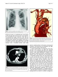
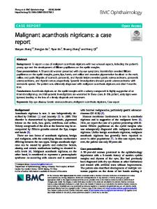
An 81-year-old man was referred for evaluation of a mediastinal mass. He visited the emergency department presenting with hemoptysis, and the mass was revealed subsequently on chest computed tomography (CT). He complained of no pain but suffered respiratory symptoms including cough and sputum. He had a history of colon cancer 7 years previously, and had been on hypertension medication for 10 years. The mass was located in the left upper thorax, with a long diameter of approximately 5.8 cm, exhibiting poor enhancement on chest CT (Fig. 1a). There was no evidence of either aortic dissection or significant steno-occlusive lesion on reconstructed CT angiography (CTA) (Fig. 1b). The clinician suspected malignancy in the mediastinum, such as a thymic carcinoma, or possible primary lung cancer and metastasis. The patient underwent FDG PET/CT to evaluate the mediastinal mass. PET/CT images were acquired on a Discovery MI (GE Healthcare, Milwaukee, WI, USA) at 60 min after injection of 2.96 MBq/kg of FDG. After CT (120 kVp; 10– 80 mA; section thickness, 3.75 mm), an emission PET was obtained from the thigh to the skull base (1.5 min per bed). PET images were reconstructed using the Bayesian penalized likelihood reconstruction algorithm with attenuationcorrected images. FDG PET/CT revealed heterogeneously incr
Data Loading...