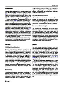
COVID-19-associated acute cerebral venous thrombosis: clinical, CT, MRI and EEG features
- PDF / 1,452,802 Bytes
- 3 Pages / 595.276 x 790.866 pts Page_size
- 36 Downloads / 317 Views
RESEARCH LETTER
Open Access
COVID-19-associated acute cerebral venous thrombosis: clinical, CT, MRI and EEG features Fabian Roy-Gash1* , De Mesmay Marine1, Devys Jean-Michel1, Vespignani Herve2, Blanc Raphael3 and Engrand Nicolas1
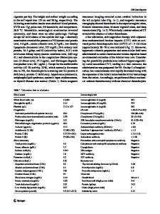
Keywords: Cerebral venous thrombosis, Intracerebral hematoma, Electroencephalography, SARS-CoV-2, COVID-19, Pandemics, Diagnostic imaging Dear editor, Many recent COVID-19 series have reported arterial or venous thrombosis (stroke, pulmonary embolism, etc.) [1, 2]. Here, we report a case of COVID-19 associated cerebral venous thrombosis (CVT) with dramatic evolution. On April 3, 2020, a 63-year-old female presented to the emergency department because of aphasia and right hemiplegia. She had a 12-day history of fever, cough, and anosmia. Her husband was hospitalized in intensive care for confirmed COVID-19 acute respiratory distress syndrome (ARDS). Brain MRI showed a large left temporal brain hemorrhage and a suspicion of CVT confirmed on a venous brain CT scan and chest CT showed typical COVID-19 patchy ground-glass opacities in both lungs (Fig. 1). The patient suddenly suffered a clinical status epilepticus and was administered i.v. lacosamide. Laboratory results showed hyperfibrinogenemia (7.2 g/L) and high ferritin levels (1427 μg/L). The nasopharyngeal and bronchial samples were negative for SARS-CoV-2.
* Correspondence: [email protected] Address for reprint: Fondation Ophtalmologique Adolphe de Rothschild, 29 Rue Manin, 75019 Paris, France. 1 Neuro-Intensive Care Unit, Fondation Ophtalmologique Adolphe de Rothschild, 29 rue Manin, 75019 Paris, France Full list of author information is available at the end of the article
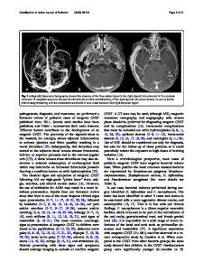
Most common causes of genetic thrombotic disorders and antiphospholipid antibody syndrome were excluded. The patient was started on an intravenous curative dose of heparin anticoagulation. Electroencephalograpy (EEG) showed background theta activity unreactive to nociceptive stimulus, with pseudo-periodic activity of a short period composed of slow di-phasic waves irradiating towards the anterior regions (Fig. 2). Although subtle status epilepticus could not be excluded, the aspect was not typical and other successive EEG traces would confirm this non-epileptic paroxystic pseudo-periodic pattern. The patient eventually underwent surgical intracranial hematoma evacuation followed by decompressive craniectomy. On April 17th, brain CT scan revealed a new intracranial contralateral bleeding most likely following contralateral venous thrombosis despite being properly treated with intravenous heparin. Venous angiography showed persistent left thrombosis (Fig. 1). On April 25th, the patient was tested positive for SARS-CoV-2 plasmatic IgG and IgM (ELISA test). On April 29th, the patient died following therapeutic limitation after ethical consultation group expertise. Although both samples were negative for SARS-CoV2, we considered the patient infected by it, given the initial symptomatology, the confirmed infection in one relative, the specific aspect of the thoracic
Data Loading...