COVID-19 in decompensated cirrhosis
- PDF / 605,205 Bytes
- 3 Pages / 595.276 x 790.866 pts Page_size
- 88 Downloads / 313 Views
LETTER TO THE EDITOR
COVID‑19 in decompensated cirrhosis Mohamed Rela1 · Vaibhav Patil1 · Gomathy Narasimhan1 · Dinesh Jothimani1 Received: 4 June 2020 / Accepted: 6 September 2020 © Asian Pacific Association for the Study of the Liver 2020
Dear Editor COVID-19 is associated with higher mortality in patients pre-existing comorbidities. There has been several reports recently describing abnormalities in liver function tests (LFT) in COVID-19 patients ranging from very mild abnormalities in the non-cirrhotic patients to moderate elevation of liver enzymes in the cirrhotic patients [1, 2]. Our recent review showed that 14–53% of patients with COVID-19 developed hepatic dysfunction and 2–11% of patients had underlying chronic liver disease [3]. In this context, we would like to report two of our patients with decompensated cirrhosis presented with acute-onchronic liver failure (ACLF) due to SARS-CoV-2 infection. Both patients had a rapidly progressive liver failure followed by respiratory failure and succumbed to the disease within days of the diagnosis. Case 1 A 69-year-old gentleman with NASH cirrhosis and chronic portal vein thrombosis was diagnosed in March 2020 with a small hepatocellular carcinoma. His bilirubin was 8.8 mg/dl, alanine amino transferase (ALT) 42 U/L, albumin 2.6 g/dL, INR 1.54 and creatinine 1.28 mg/dl. His Child Pugh score was 10 (Class C) and his model for end-stage liver disease (MELD) was 23. His HBsAg, antiHBcAb, and anti-HCV antibodies were negative. He was being worked up for liver transplantation. He presented again in mid-April with 5 days history of abdominal distension, altered sensorium and breathing difficulty. His past medical history includes type 2 diabetes mellitus and hypertension. Clinical examination revealed jaundice, moderate ascites (grade 2), pedal edema and grade 2 encephalopathy. His respiratory rate was 28 per minute and oxygen saturation 92% on room air. His arterial blood gas showed pH 7·314, pO2 55·4 mm Hg and p CO2 36·8 mmHg. His Hb 12.1 g/dl, * Dinesh Jothimani [email protected] 1
Institute of Liver Disease and Transplantation, Dr Rela Institute and Medical Centre, Bharath Institute for Higher Education and Research, Chennai, India
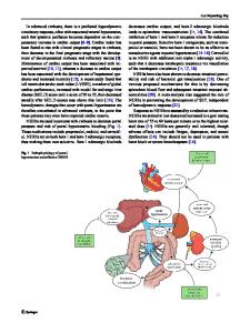
white blood cell count 7780 cells/mm3, differential count showed Neutrophils 6690 cells/mm3 (86%), lymphocytes 311 cells/mm3 (4.0%), and platelet count 139 × 109/L. He was negative for Hepatitis A and Hepatitis E IgM antibodies. His serum bilirubin was 20·2 mg/dl, ALT 58 U/L, albumin 2.7 g/dL, International normalized ratio (INR) 2.82 and creatinine was 2·83 mg/dl (not on diuretics). His Child Pugh score was 13 (Class C) and MELD was 39. The marker of acute phase reaction such as C-Reactive protein (CRP) was not available. His chest X-ray showed left-sided pleural effusion. A CT thorax revealed bilateral patchy ground glass attenuation of the lung parenchyma (Fig. 1a). His nasopharyngeal swab for SARS-CoV-2 RT-PCR was positive. Ascitic fluid analysis did not show spontaneous bacterial peritonitis. He was comme
Data Loading...