CT-guided hook-wire localization of malignant pulmonary nodules for video assisted thoracoscopic surgery
- PDF / 5,778,870 Bytes
- 7 Pages / 595.276 x 790.866 pts Page_size
- 65 Downloads / 285 Views
(2020) 15:307
RESEARCH ARTICLE
Open Access
CT-guided hook-wire localization of malignant pulmonary nodules for video assisted thoracoscopic surgery Huijun Zhang1*†, Ying Li2†, Nadier Yimin1, Zelai He3* and Xiaofeng Chen1*
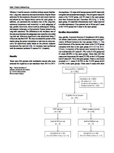
Abstract Objectives: Video assisted thoracoscopic surgery (VATS) can currently be used to diagnose and treat pulmonary nodules. However, intraoperative location of pulmonary nodules in VATS is challenging due to their small diameter and deep location in the pulmonary parenchyma. The purpose of this study was to report the clinical safety and effectiveness of CT-guided hook-wire for preoperative localization of malignant pulmonary nodules smaller than 1 cm in diameter. Methods: From February 2017 to January 2018, we collected the data of 80 patients with malignant pulmonary nodules less than 1 cm in diameter who underwent CT-guided hook-wire preoperative localization and VATS surgery. The effectiveness of preoperative localization was evaluated based on surgical duration, success rate of VATS surgery, and localization-related complications. Results: The diameter of pulmonary nodules were 0.85 ± 0.17 mm with a distance to the pleural surface of 19.66 ± 14.10 mm. The length of the hook-wire in the lung parenchyma was 29.17 ± 13.14 mm and hook-wire dislodgement occurred in 2 patients. Complications included 27 cases of minor pneumothorax and 18 cases of mild parenchymal hemorrhage. A significant correlation was observed between the length of the hook-wire in the lung parenchyma and mild parenchymal hemorrhage (P = 0.044). The average time of hook-wire localization was 9.0 ± 2.6 min and the average operation time for VATS was 89.02 ± 23.35 min without conversion thoracotomy. Conclusions: CT-guided hook-wire localization of the lesion during VATS resection is safe for malignant pulmonary nodules with diameter less than 1 cm. Keywords: Malignant pulmonary nodules, Hook-wire localization, Video assisted thoracoscopic surgery
Introduction Low-dose spiral computed tomography (CT) screening for early lung cancer is a promising strategy for improving lung cancer survival and can reduce lung cancer * Correspondence: [email protected]; [email protected]; [email protected] † Huijun Zhang and Ying Li contributed equally to this work. 1 Department of Cardiothoracic Surgery, Huashan Hospital of Fudan University, Shanghai 200040, China 3 Department of Radiation Oncology, The First Affiliated Hospital of Bengbu Medical College, Bengbu 233000, China Full list of author information is available at the end of the article
deaths by 20% [1]. Due to the widespread use of lowdose spiral CT, an increasing number of pulmonary nodules have been detected in the early stages of lung cancer, especially those featuring ground-glass opacity (GGO). Of these nodules, 59–73% are malignant, therefore rapid and accurate histological diagnosis and treatment are necessary [2]. The effectiveness of routine procedures such as transbronchial or CT-guided fineneedle aspiration biopsy may be limited by the small diameter of the pulmona
Data Loading...