Diffuse Leptomeningeal Glioneuronal Tumors: Histology. Is It a New Entity?
The updated World Health Organization (WHO) Classification of Tumors of the Central Nervous Systems has recently expanded the category of mixed glioneuronal tumors with three new entities (i.e. papillary glioneuronal tumor, rosetted glioneuronal tumor wit
- PDF / 520,298 Bytes
- 7 Pages / 504.57 x 720 pts Page_size
- 55 Downloads / 302 Views
13
Marina P. Gardiman and Matteo Fassan
Contents
Abstract
Introduction ............................................................
102
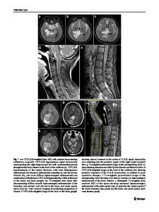
Clinical and Neuroradiological Findings .............
102
Pathological Findings.............................................
103
Discussion................................................................
105
Conclusions .............................................................
106
References ...............................................................
107
M.P. Gardiman (*) Surgical Pathology Unit, Padua University Hospital, Via Giustiniani 1, 35121 Padua, Italy e-mail: [email protected] M. Fassan Surgical Pathology Unit, Padua University Hospital, Via Gabelli, 61, 35121 Padua, Italy
The updated World Health Organization (WHO) Classification of Tumors of the Central Nervous Systems has recently expanded the category of mixed glioneuronal tumors with three new entities (i.e. papillary glioneuronal tumor, rosetted glioneuronal tumor with neuropil-like islands, and rosette-forming glioneuronal tumor of the fourth ventricle). This classificatory extension is in part the consequence of the constantly increasing availability of novel immunostains, which have enabled the pathologists’ community to more readily identify neuronal differentiation in tumors morphologically resembling glial neoplasms. However, despite this growing list of new entities, in the routine diagnostic practice it is still possible to encounter glioneuronal tumors that cannot be placed into any of the well-defined WHO categories. We have recently reported the unusual radiologic and pathologic findings of four analogous paediatric cases in which both the morphological and the immunohistochemical findings strongly supported a glioneuronal commitment of the tumors. Because of the unique overlapping clinical and neuroradiological characteristics, we propose to consider this group of neoplasms as a new possible distinct pathological and clinical entity in the group of glioneuronal tumors.
M.A. Hayat (ed.), Tumors of the Central Nervous System, Volume 9, DOI 10.1007/978-94-007-5488-1_13, © Springer Science+Business Media Dordrecht 2012
101
102
Introduction Glioneuronal tumors are a group of primary brain neoplasms of relatively recent acquisition in the World Health Organization (WHO) Classification of the Central Nervous System (CNS) tumors. This classification has recently been expanded with new recognized entities such as rosetteforming tumor of the fourth ventricle, papillary glioneuronal tumor and rosetted glioneuronal tumor/glioneuronal tumor with neuropil-like islands (Louis et al. 2007). Glioneuronal tumors are characterized by a biphasic neurocytic and glial population. The neuronal component consists of synaptophysin-positive neurocytes with round nuclei and clear cytoplasm occasionally intermingled with neurons and intermediatesized “ganglioid” cells, whereas the glial component exhibits features of glial fibrillary acidic protein (GFAP) positive astrocytes. The his
Data Loading...