Dose distribution of intensity-modulated proton therapy with and without a multi-leaf collimator for the treatment of ma
- PDF / 1,166,253 Bytes
- 10 Pages / 595.276 x 790.866 pts Page_size
- 48 Downloads / 321 Views
RESEARCH
Open Access
Dose distribution of intensity-modulated proton therapy with and without a multileaf collimator for the treatment of maxillary sinus cancer: a comparative effectiveness study Soichi Sugiyama1,2, Kuniaki Katsui3* , Yuki Tominaga4, Takahiro Waki2, Norihisa Katayama5, Hidenobu Matsuzaki6, Shin Kariya7, Masahiro Kuroda8, Kazunori Nishizaki7 and Susumu Kanazawa1
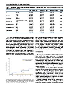
Abstract Background: Severe complications, such as eye damage and dysfunciton of salivary glands, have been reported after radiotherapy among patients with head and neck cancer. Complications such as visual impairment have also been reported after proton therapy with pencil beam scanning (PBS). In the case of PBS, collimation can sharpen the penumbra towards surrounding normal tissue in the low energy region of the proton beam. In the current study, we examined how much the dose to the normal tissue was reduced by when intensity-modulated proton therapy (IMPT) was performed using a multi-leaf collimator (MLC) for patients with maxillary sinus cancer. Methods: Computed tomography findings of 26 consecutive patients who received photon therapy at Okayama University Hospital were used in this study. We compared D2% of the region of interest (ROI; ROI-D2%) and the mean dose of ROI (ROI-mean) with and without the use of an MLC. The organs at risk (OARs) were the posterior retina, lacrimal gland, eyeball, and parotid gland. IMPT was performed for all patients. The spot size was approximately 5–6 mm at the isocenter. The collimator margin was calculated by enlarging the maximum outline of the target from the beam’s eye view and setting the margin to 6 mm. All plans were optimized with the same parameters. Results: The mean of ROI-D2% for the ipsilateral optic nerve was significantly reduced by 0.48 Gy, and the mean of ROI-mean for the ipsilateral optic nerve was significantly reduced by 1.04 Gy. The mean of ROI-mean to the optic chiasm was significantly reduced by 0.70 Gy. The dose to most OARs and the planning at risk volumes were also reduced. Conclusions: Compared with the plan involving IMPT without an MLC, in the dose plan involving IMPT using an MLC for maxillary sinus cancer, the dose to the optic nerve and optic chiasm were significantly reduced, as measured by the ROI-D2% and the ROI-mean. These findings demonstrate that the use of an MLC during IMPT for maxillary sinus cancer may be useful for preserving vision and preventing complications. Keywords: Multi-leaf collimator, Chemoradiotherapy, Intensity-modulated proton therapy, Pencil beam scanning, Maxillary sinus cancer
* Correspondence: [email protected] 3 Departments of Proton Beam Therapy, Dentistry and Pharmaceutical Science, Okayama University Graduate School of Medicine, 2-5-1 Shikata-cho, Kita-ku, Okayama 700-8558, Japan Full list of author information is available at the end of the article © The Author(s). 2019 Open Access This article is distributed under the terms of the Creative Commons Attribution 4.0 International License (http://creativecommons.org/licenses/by
Data Loading...