Elderly Japanese Standard Data of Echocardiography; From J-LONG study
- PDF / 568,680 Bytes
- 8 Pages / 595.276 x 790.866 pts Page_size
- 67 Downloads / 301 Views
ORIGINAL INVESTIGATION
Elderly Japanese Standard Data of Echocardiography; From J‑LONG study Yoshihiro Seo1 · Tomoko Ishizu2 · Masaki Ieda2 · Nobuyuki Ohte1 on behalf of J-LONG Study Investigators Received: 24 December 2019 / Revised: 26 February 2020 / Accepted: 5 April 2020 © Japanese Society of Echocardiography 2020
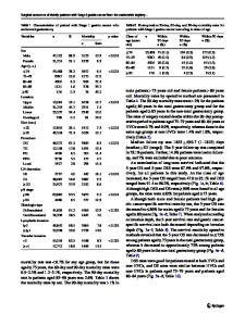
Abstract Background Age-related changes in cardiac morphology and function have not been unknown in the elderly. The Japanese eLderly data Of Normal echocardioGraphy (J-LONG) study is a prospective multicenter cohort study and aimed to investigate the echocardiographic data in the healthy Japanese elderly. Methods Thirty domestic facilities participated in this study, and 130 healthy subjects (57 men, 73 women, 79.6 ± 4.7 years, range 75–98 years, interquartile range 76–82 years) were enrolled in this cohort. Echocardiographic and clinical data sets were obtained in each facility, and total analyses were performed in the University of Tsukuba. Results Almost all cardiac morphological data were significantly larger in men than those in women. However, corrected data by body surface area (BSA) were similar or closer between genders. As a gender difference, the negative correlation between BSA and age was observed in women only (r = − 0.46, p 50% thicker than the thickness of the septum at its mid-distal-point [6]. AVS was identified by aortic cusp thickening, normal aortic cusp excursion, and peak transaortic valve flow velocity less than 2.0 m/s. MAC was defined by an intense echocardiograph-producing structure located at the junction of the atrioventricular groove and posterior mitral leaflet [7]. Severity was qualitatively determined in parasternal short-axis view at the level of the mitral annulus as mild (focal, limited increase in echo-density of the mitral annulus), moderate (marked echodensity involving one-third to one-half of the ring circumference), or severe (marked echo-density involving more than one-half of the circumference of the ring or with intrusion into the LV inflow tract). Comprehensive echocardiographic studies for both the left and right sides of the heart were performed according to established guidelines [8]. LV end-diastolic diameter (LVDd), thickness of interventricular septum (IVST), posterior wall thickness (PWTd), and end-systolic diameter (LVDs) were measured at parasternal long-axis views at mitral tip level. In patients with sigmoid septum, the parameters were measured at a distal site of septal hypertrophy. LV end-diastolic volume (LVEDV), LV endsystolic volume (LVESV), and left ventricular ejection fraction (LVEF) were measured by the biplane Simpson’s
Journal of Echocardiography
method from the apical 4-chamber and 2-chamber views. Relative wall thickness was estimated as 2 × (diastolic LV posterior wall thickness)/LV end-diastolic diameter. LV mass (LVM) was calculated using Devereux’s formula. Maximum left atrial volume (LAV) was measured by the biplane Simpson’s method. LVDd, LVDs, LVEDV, LVESV, LVM and LAV were indexed to body surface area (LVDdI, LVD
Data Loading...