Electrochemical-microscopy analysis of bio-functionalized diamond surfaces
- PDF / 1,068,843 Bytes
- 6 Pages / 612 x 792 pts (letter) Page_size
- 49 Downloads / 249 Views
1039-P07-03
Electrochemical-microscopy analysis of bio-functionalized diamond surfaces Hiroshi Uetsuka, Nianjun Yang, Norio Tokuda, and Christoph E. Nebel National Institute of Advanced Industrial Science and Technology, Tsukuba, 305-8568, Japan
ABSTRACT DNA sensors fabricated on single-crystalline B-doped diamond (SBDD) electrodes were characterized by scanning electrochemical microscopy (SECM). These experiments show that circular arranged DNA sensors of typically 2 mm diameter show some spatial variations in electrochemical response. The variation of the diamond electrode with respect to DNA bonding is electrochemically characterized and discussed in the context of ion attraction/repulsion by the negatively charged backbones of DNA and the negatively charged diamond surface. Our results show that repulsive forces affect the mediator propagation of Fe(CN)64- in the close vicinity of the DNA layer which can be used to investigate DNA density variations on sensor areas.
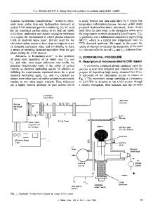
INTRODUCTION Great attention has been attracted by bio-sensor devices based on diamond because diamond is known to be biocompatibility, chemical inert, shows excellent electrochemical properties and long term chemical stability of bio-molecules bonded to it [1]. Electrochemical DNA sensors consisting of a phenyl layer, a cross-linker layer, and a DNA layer have been studied in detail macroscopically [2,3]. From these experiments it became clear that optimization of each of these layers is mandatory for excellent sensor performance [4,5]. Conventional electrochemical analysis applies in most cases a macroscopic working electrode as sensor which averages the electrochemical response of a large area. Diamond itself can be laterally inhomogeneous due to some doping variations, due to grain boundaries, and surface defect variations. Electrochemical bonding of phenyl molecules to diamond will most likely enhance these inhomogeneities as attachment will preferentially take place on areas with good electrochemical interface while on areas where no current is flowing the grafted will be suppressed. Up-to-now, a microscopic analysis of sensors is missing. In this paper, we report about the application of scanning electrochemical microscopy (SECM) where a small electrode of typically 10 µm in diameter is used as working electrode. This allows to characterize the electrochemical properties with high lateral resolution. Twodimensional (2D) mapping of electrochemical activity of electrodes can be applied. In this study, SECM was used to investigate the electrochemical homogeneity of DNA sensors fabricated on atomically smooth B-doped diamond surfaces.
EXPERIMENTAL DETAILS Boron-doped single-crystalline diamond films have been grown homoepitaxially on synthetic (100) Ib diamond substrates with 4 mm × 4 mm × 0.4 mm size, using microwave plasma-assisted chemical vapor deposition (CVD). The surface of these films is very smooth with a surface roughness (RMS) below 1 Å [3]. Because of ultra high doping of diamond with 3 × 1020 cm-3 boron acceptors the film shows a
Data Loading...