Endocytosis and exocytosis processes of gold nanoparticle with erythrocyte ghosts
- PDF / 476,481 Bytes
- 4 Pages / 432 x 648 pts Page_size
- 49 Downloads / 318 Views
MRS Advances © 2020 Materials Research Society DOI: 10.1557/adv.2020.299
Endocytosis and exocytosis processes of gold nanoparticle with erythrocyte ghosts Víctor Gómez Flores1 1
Institute of engineering and Technology University Autonomous of Ciudad Juarez, Ave. Del Charro #610.Col Partido Romero, Ciudad Juárez, México ZIP 32310
ABSTRACT
The interaction of spherical gold nanoparticles (AuNPs) of 20nm elaborated by Turkevich method with the erythrocytes ghosts (7-8 μm) cell membrane was evaluated. The AuNPsMembrane interaction was determined using confocal microscope, Uv-Vis spectroscopy and SEM analysis. The result show that nanoparticles larger than 20nm are adhered to the erythrocyte ghost membrane due their size and surface modification. Smaller AuNPs enter onto the cell by simple diffusion through the plasmatic membrane voids, these data may favor the best design and application in the treatments applied in biomedicine.
INTRODUCTION The use of nanoparticles in biomedical applications depends on their morphological structure and mainly on the surface properties. Currently, there is limited knowledge of the interaction of these nanoparticles with cells, specifically the effect of physical and chemical modification on the endocytosis and exocytosis [1]. Understanding this mechanism will allow better applications in drug delivery and nanomedicine area [2,3]. The most recent study carried out by Brenner and collaborators [4], used erythrocytes as nanocarriers, for the release of nanoparticles and to check their accumulation in the different organs exposed ex vivo, demonstrating that high doses of nanoparticles can enter erythrocytes, using two kinds of nanoparticles, 1) polystyrene and 2) nanogel. Currently Xing et al.[5] conducted a study for a new drug delivery system based on gold nanoparticles coated with liposomes, where doxorubicin encapsulated an anticancer drug. 1
Downloaded from https://www.cambridge.org/core. UCSD University of California San Diego, on 13 Jul 2020 at 20:13:07, subject to the Cambridge Core terms of use, available at https://www.cambridge.org/core/terms. https://doi.org/10.1557/adv.2020.299
In the present study, 20 nm diameter AuNPs, arranged on the erythrocyte ghosts membrane, were used to evaluate the interaction of AuNPs (characterized with Uv-Vis and scanning electron microscope) and the cell membrane determining the interaction between both.
EXPERIMENTAL DETAILS Synthesis of AuNPs The gold nanoparticles were synthesized by the Turkevich method [6]. Two precursor solutions were used: 18ml of chloroauric acid (HAuCl4, 200mg / dL 0.5mM, Sigma Aldrich®) and 2 ml of Sodium Citrate (Na3C6H5O7, JT Baker®). HAuCl4 was first heated under magnetic stirring to boiling, at which point the 2 ml of sodium citrate was added over 30 minutes. There was a change in color from crystalline to ruby red, which showed the formation of nanoparticles. Then the solution was allowed to cool to room temperature for later use.
Erythrocyte ghosts The method of Niggli et al [7] was used, with some modifica
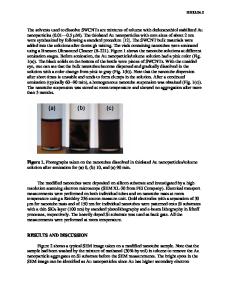
Data Loading...