Esogastro-Pleuro-Bronchial Fistula: an Unusual Complication After Sleeve Gastrectomy
- PDF / 159,761 Bytes
- 2 Pages / 595.276 x 790.866 pts Page_size
- 95 Downloads / 298 Views
LETTER TO THE EDITOR
Esogastro-Pleuro-Bronchial Fistula: an Unusual Complication After Sleeve Gastrectomy Jesus Badia-Closa 1
&
Alexis Luna 1 & Sandra Montmany 1 & Pere Rebasa 1 & Salvador Navarro 1
# Springer Science+Business Media, LLC, part of Springer Nature 2020
Bariatric surgery has become one of the main treatments for morbid obesity. Sleeve gastrectomy (SG) is among the most performed surgical techniques worldwide, surpassing Roux-en-Y Gastric By-Pass (RYGBP) as the most popular procedure in recent years 1. Regarding its safety, the main limitation of this technique is gastric fistulae (GF). What causes them is still unclear, but an increase in intragastric pressure and a modification in the vascularization after surgery can lead to a leak (usually in the upper part of the staple line, closer to the gastroesophageal junction) 2, hence developing a GF. Most GF can be safely managed with conservative treatment or with endoscopic or minimally invasive surgical procedures, but in some cases, these are not sufficient and a more radical approach is needed [1]. Acute GF (AGF) incidence ranges between 1 and 3% 3 after SG, while Chronic GF (CGF) is much lower at 0.1% 4, 5 after SG. A particular kind of CGF is the one that also affects the pleura or even the lungs, developing an esogastrobronchopleural fistula (EGBPF). Among the available treatments for CGF, there are endoscopic and surgical options, such as Roux-en-Y fistulojejunostomy 6, conversion to a RYGBP or total gastrectomy 1. If the pleura and/or lungs are affected, it may require lung or diaphragmatic resections. We present a patient who developed an EGBPF after SG and the treatment she underwent. A 56-year-old woman who underwent a SG (open surgery due to a large incisional hernia from a previous surgery that was repaired in the same operation) for class II obesity (BMI * Jesus Badia-Closa [email protected] 1
Gastrointestinal Unit, Department of General and Digestive Surgery, Parc Taulí University Hospital, Universitat Autònoma de Barcelona (UAB), Parc Taulí s/n, 08208, Sabadell, Barcelona, Spain
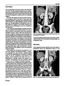
36.7) associated with high blood pressure and type-2 diabetes. One year after surgery, she was readmitted to the hospital due to a subphrenic abscess caused by a proximal GF. It was treated with antibiotics and she was discharged a week later. For personal reasons, the patient was lost in follow-up. Eleven years later, she was admitted to the emergency room with fever and hemoptysis. The abdominal CT showed a left subphrenic abscess, also affecting the pleura, and with the contrast administered by endoscopy seen inside the bronchium (EGBPF) (Figure 1). She was initially treated with broad-spectrum antibiotics and a trans-gastric drainage of the abscess, but the fever persisted. An endoluminal vacuum therapy device (Endosponge©) was used, with no improvement, so the patient was programmed for elective surgery. Through a midline incision, the subphrenic space was accessed and the abscess debrided. The fistula was identified and removed with a stapler. A gastric po
Data Loading...