Expression of programmed death-ligand 1 (PD-L1) in human pituitary neuroendocrine tumor
- PDF / 1,129,362 Bytes
- 9 Pages / 595.276 x 790.866 pts Page_size
- 107 Downloads / 287 Views
ORIGINAL ARTICLE
Expression of programmed death‑ligand 1 (PD‑L1) in human pituitary neuroendocrine tumor Valentine Suteau1,2 · Alexandre Collin3 · Philippe Menei4,5 · Patrice Rodien1,2,5 · Marie‑Christine Rousselet3,5 · Claire Briet1,2,5 Received: 6 January 2020 / Accepted: 13 May 2020 © Springer-Verlag GmbH Germany, part of Springer Nature 2020
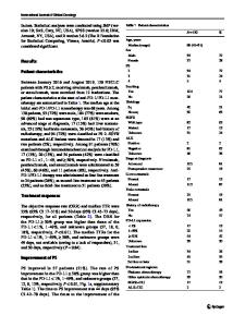
Abstract Objective To explore the programmed death-ligand 1 (PD-L1) expression in varied subtypes of pituitary neuroendocrine tumors with assessment of their clinical behavior at diagnosis and follow-up. Methods We conducted a retrospective monocentric study, including all patients operated in the Academic Hospital of Angers (France) for a pituitary neuroendocrine tumor between 2012 and 2018. PDL-1 immunostaining was performed using a European Conformity—In Vitro Diagnostic-labeled anti-PDL1 antibody (clone 22C3). PD-L1 immunostaining was evaluated as the percentage of tumor cells showing positive membrane staining, into four grades: grade 0 = 2 mitoses per 10 high-power field, Ki-67 ≥ 3%, p53 positive
c
Stratification of the tumors into four grades: grade 1a, noninvasive; grade 1b, noninvasive and proliferative; grade 2a, invasive; grade 2b, invasive and proliferative; missing data for one patients
We explored the prognostic impact of PD-L1 expression in PitNET according to HYPOPRONOS classification. Invasion, defined as cavernous and/or sphenoid sinus invasion (CSI) on MRI, was not more frequent in the PD-L1-positive group. Sato et al. recently reported a trend to higher, though not significant PD-L1 expression in PitNET with cavernous sinus invasion (8/17 CSI + versus 1/10 CSI- with PD-L1 > 5%) [18]. Regarding the proliferative activity, we found that only one of 17 tumors with high Ki-67 (≥ 3%) and/or positive p53 expression was PD-L1 positive. No significant differences could be established for any proliferation criteria, and this may be due to the small number of PDL1-positive cases. When we classified the tumors according to HYPOPRONOS grade, PD-L1-positive tumors were never classified in grade 1b or 2b, suggesting that PD-L1 expression seemed to be associated with less proliferation. Divergent results were previously observed about Ki-67 and PD-L1 expression, thus leaving this question open [15, 17]. We also compared the evolution of PD-L1 expression in 6 patients with recurrence. Neither primary tumors
13
displayed PD-L1 expression nor the relapse tissue, which is inconsistent with the results of Mei et al. [15]. Therefore, PitNET progression in our study was not accompanied by PD-L1 expression. However, PitNET are benign tumor with sometimes a local aggressive behavior, unlike carcinoma, which is malignant tumor. The description in the literature of the spectacular response of one case of ACTH-PitNET carcinoma with ipilimumab and nivolumab treatment is puzzling in regard to cumulated data concerning PD-L1 in PitNET. However, no PD-L1 expression was demonstrated in the liver metastasis, which may suggest that ipilimumab (anti CTLA-4) rather than nivolum
Data Loading...