Fabrication and Characterization of MEMS-Based Structures from a Bio-Inspired, Chemo-Responsive Polymer Nanocomposite
- PDF / 293,760 Bytes
- 6 Pages / 432 x 648 pts Page_size
- 87 Downloads / 476 Views
Fabrication and Characterization of MEMS-Based Structures from a Bio-Inspired, Chemo-Responsive Polymer Nanocomposite Allison E. Hess1and Christian A. Zorman1,2 Department of Electrical Engineering and Computer Science, Case Western Reserve University, Cleveland, OH, 44106 2 Advanced Platform Technology Center of Excellence, Louis Stokes Cleveland VA Medical Center, Cleveland, OH, 44106
1
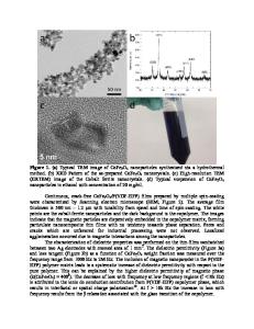
ABSTRACT This paper reports the development of micromachining processes, as well as electrical and mechanical evaluation of a stimuli-responsive, mechanically-dynamic polymer nanocomposite for biomedical microsystems. The nanocomposite, which consists of a cellulose nanofiber network embedded in a poly(vinyl acetate) matrix, was shown to display a switchable stiffness comparable to bulk samples, with a Young’s modulus of 3570 MPa in the dry state, which reduced to ~25 MPa in the wet state, with a stiff-to-flexible transition-time dependent on exposed surface area. Upon immersion in phosphate buffered saline, the ac resistance through the PVAc-TW thickness was found to reduce from 8.04 MΩ to ~17 kΩ. Electrochemical impedance of an Au electrode on PVAc-TW was found to be ~178 kΩ at 1 kHz, and this was found to be stable as the probe shank was flexed to compress the metal, but increased with increasing flex angle when the metal was flexed into a tensile state. INTRODUCTION Long-term (>20 years) viability of intracortical probes is essential for advancing clinical applications of neural recording technology for improving the quality of life for people with neurological conditions, such as spinal cord injury or Parkinson’s disease. However, current probes, which are primarily based on Si,1, 2 are often unable to record neural signals after a few months due to glial scarring that electrically and mechanically isolates electrodes from neurons.3 It has been hypothesized that strain induced by micromotion between the stiff Si (Young’s modulus, E=~160 GPa) and soft cortical tissue (E=~10 kPa) triggers the immune response that produces this cellular sheath,4 and that a probe based on a material that better matches cortical tissue mechanics would alleviate the issue, allowing for long-term electrode viability. In response, polymer-based intracortical probes using polyimide5 and parylene6 have been developed, but have suffered from the inability to penetrate the pia mater without buckling unless stiffened with a rigid backbone or gel-filled microfluidic channel. A stimuli-responsive polymer nanocomposite, inspired by the switchable stiffness behavior of the sea cucumber (Cucumaria frondosa) dermis,7 has recently been developed by our colleagues for use as a substrate for adaptive intracortical probes for potentially long-term neural recording.8 Comprised of a soft poly(vinyl acetate) matrix with rod-like cellulose nanofiber fillers (also referred to as whiskers) obtained from filter-feeding sea creatures known as tunicates, the stiffness of the polymer nanocomposite (PVAc-TW) is modulated by interactions between the surface hydroxyl groups on
Data Loading...