Hemoglobin Expression in Neurons and Glia After Intracerebral Hemorrhage
The purpose of this study was to examine the expression of hemoglobin (Hb) in the brain after intracerebral hemorrhage (ICH) and the effects of hemin and iron on neuronal Hb.
- PDF / 214,752 Bytes
- 5 Pages / 595.276 x 790.866 pts Page_size
- 89 Downloads / 337 Views
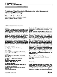
Abstract The purpose of this study was to examine the expression of hemoglobin (Hb) in the brain after intracerebral hemorrhage (ICH) and the effects of hemin and iron on neuronal Hb. For the in vivo studies, male Sprague-Dawley rats received either a sham operation or an ICH. The rats were killed 1, 4, 24 or 72 h later, and brains were used for real-time polymerase chain reaction (PCR) and immunohistochemistry. For the in vitro study, primary cultured neurons were exposed to either hemin or vehicle. Some neurons also received treatment with deferoxamine, an iron chelator. Neurons were collected 24 h later for real-time PCR. We found that a-globin (HbA) and b-globin (HbB) mRNA levels in the ipsilateral basal ganglia are significantly increased after ICH, and Hb is localized in neurons and glia cells. Exposure of neurons to hemin also upregulated HbA and HbB mRNA levels. Hemin-induced HbA and HbB expression in cultured neurons was reduced by deferoxamine treatment. These results indicate that ICH increases HbA and HbB expression in neurons and glia cells, and that heme and iron may be important factors in inducing endogenous Hb expression after ICH. Keywords Intracerebral hemorrhage · Hemoglobin · Hemin · Iron · Deferxomine
Y. He, Y. Hua, R.F. Keep, and W. Liu Department of Neurosurgery, University of Michigan, Ann Arbor, MI, USA M.M. Wang Department of Neurology, University of Michigan, Ann Arbor, MI, USA G. Xi (*) Department of Neurosurgery, University of Michigan, Ann Arbor, MI, USA and Department of Neurosurgery, University of Michigan, R5018 Biomedical Science Research Building, Ann Arbor, 48109-2200, MI, USA e-mail: [email protected]
Introduction Vertebrate hemoglobin (Hb) is a heterotetramer of two a-globin (HbA) and two b-globin (HbB) subunits. Each subunit contains a hydrophobic pocket in which a heme molecule binds tightly and allows for the reversible binding of oxygen [1]. Vertebrate Hb has been traditionally thought to be exclusively present in cells of erythroid lineage. However, recent studies have shown that Hb is expressed in a variety of nonhematopoietic tissues and cell lines, including macrophages, epithelial cells and mesangial cells [2–4]. The presence of Hb in neurons has also been reported, and neuronal Hb is inducible after cerebral and ischemic preconditioning [5–8]. Intracerebral hemorrhage (ICH) is a common and often fatal subtype of stroke. After ICH, erythrocyte lysis can happen in the early phase, and Hb is released from the lysed blood cells [9]. There is evidence suggesting that free Hb is deleterious to the brain after ICH. Exogenous Hb can cause brain injury when injected into the brain in vivo [10, 11] or added to cerebral cortical neurons in vitro [12, 13]. Currently, the effects of ICH on endogenous Hb expression are unknown. In this study, we examined the change of endogenous Hb expression after ICH. The effects of hemin and iron on neuronal Hb expression were also examined.
Materials and Methods Animal Preparation and Intracerebral Infusion The University of Michiga
Data Loading...