Heterotropic Gastric Tissue in Duodenal Bulb Mimicking Duodenal Cancer: An Unusual Duodenal Lesion
- PDF / 197,181 Bytes
- 2 Pages / 595.276 x 790.866 pts Page_size
- 80 Downloads / 286 Views
LETTER TO THE EDITOR
Heterotropic Gastric Tissue in Duodenal Bulb Mimicking Duodenal Cancer: An Unusual Duodenal Lesion Rizwan Kibria & Shazdeh Butt & Syed A. Ali & Salma Akram
Published online: 6 November 2009 # Humana Press Inc. 2009
Keywords heterotropic gastric mucosa . gastric metaplasia . duodenal polyp
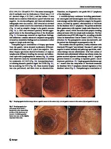
To the Editor, A 50-year-old man with no known prior medical problems underwent an esophagogastroduodenoscopy for dysphagia. The esophagus and the stomach appeared to be normal. Biopsies were obtained from the mid- and the distal esophagus to r/o eosinophilic esophagitis (EE). In the duodenal bulb, an irregular, sessile polypoid lesion, 1.5 cm in size, was noted on the anterior wall, which was also biopsied. The color of the lesion’s surface was indistinguishable from that of the normal duodenal mucosa surrounding the lesion (Figs. 1, 2). No other lesions were noted in the duodenum. Esophageal biopsies were negative for EE, and the duodenal polyp biopsies showed welldifferentiated, mature gastric glands with chief cells, parietal cells, and mucus cells. Giemsa stain was negative for Helicobacter pylori, and no metaplasia or abnormal cells were observed (Figs. 3, 4). Based on these results, a diagnosis of heterotropic gastric tissue in the duodenal bulb (HGTDB) was made. An upper gastrointestinal series and a computed tomography of abdomen were unremarkable. Manometry was done to further investigate his dysphagia which revealed nonspecific esophageal motility disorder. He was subsequently treated with a calcium channel blocker with a good response to treatment. He has so far completed 1-year follow-up and remains asymptomatic. R. Kibria (*) : S. Butt : S. A. Ali : S. Akram School of Medicine, Wright State University, Dayton, OH, USA e-mail: [email protected]
Ectopic tissue containing gastric epithelium with parietal cells may be found in the esophagus, and rarely, adenocarcinomas arise in these foci. However, HGTDB is a rare congenital lesion [1]. In the past, the term heterotopia was used interchangeably with gastric metaplasia (GM), which is an acquired lesion [2]. Histologically, HGTDB is characterized by the presence of well-differentiated gastric glands with chief and parietal cells. HGTDB is a congenital macroscopic entity that is usually found incidentally on endoscopy and has no association with H. pylori, hyperchlorhydria, duodenal ulcers, or carcinoma which is in contrast with GM. With increasing number of endoscopies being performed, physicians must differentiate HGTDB from GM, as the two conditions have entirely different clinical implications [3].
Fig. 1 Endoscopic view showing an irregular, sessile polypoid lesion on the anterior wall of duodenal bulb
J Gastrointest Canc (2009) 40:142–143
143
Fig. 4 Magnification (400×) showing well-differentiated, mature gastric glands with chief cells, parietal cells, and mucus cells Fig. 2 Endoscopic view showing an irregular, sessile polypoid lesion on the anterior wall of duodenal bulb
References
Fig. 3 Magnification (200×) showing well-differentia
Data Loading...