Histopathologic response after hydrophilic polyethylene glycol-coating stent and hydrophobic octadecylthiol-coating sten
- PDF / 1,126,773 Bytes
- 8 Pages / 595.276 x 790.866 pts Page_size
- 50 Downloads / 348 Views
BIOCOMPATIBILITY STUDIES Original Research
Histopathologic response after hydrophilic polyethylene glycol-coating stent and hydrophobic octadecylthiol-coating stent implantations in porcine coronary restenosis model Jeong Cheon Choe1 Jong Ha Park1 Han Cheol Lee1 Tae Sik Park1 Jinhee Ahn1 Jin Sup Park1 Hye Won Lee1 Jun-Hyok Oh1 Jung Hyun Choi1 Kwang Soo Cha1 Changyong Yim2 Sangmin Jeon2 ●
●
●
●
●
●
●
●
●
●
●
1234567890();,:
1234567890();,:
Received: 7 December 2019 / Revised: 7 December 2019 / Accepted: 27 October 2020 © Springer Science+Business Media, LLC, part of Springer Nature 2020
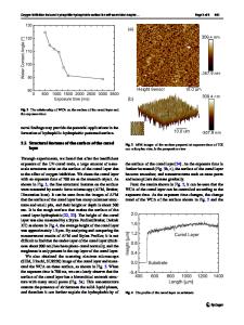
Abstract Device-related problems of drug-eluting stents, including stent thrombosis related to antiproliferative drugs and polymers, can cause adverse events such as inflammation and neointimal hyperplasia. Stent surface modification, wherein the drug and polymer are not required, may overcome these problems. We developed hydrophilic polyethylene glycol (PEG)-coating and hydrophobic octadecylthiol (ODT)-coating stents without a drug and polymer and evaluated their histopathologic response in a porcine coronary restenosis model. PEG-coating stents (n = 12), bare-metal stents (BMS) (n = 12), and ODT-coating stents (n = 10) were implanted with oversizing in 34 porcine coronary arteries. Four weeks later, the histopathologic response, arterial injury, inflammation, and fibrin scores were analyzed. A p value < 0.05 was considered statistically significant. There were significant differences in the internal elastic lamina area, lumen area, neointimal area, percent area of stenosis, arterial injury score, inflammation score, and fibrin score among the groups. Compared to the BMS or ODT-coating stent group, the PEG-coating stent group had significantly increased internal elastic lamina and lumen area (all p < 0.001) and decreased neointimal area and percent area of stenosis (BMS: p = 0.03 and p < 0.001, respectively; ODT-coating: p = 0.013 and p < 0.001, respectively). Similarly, the PEG-coating group showed significantly lower inflammation and fibrin scores than the BMS or ODT-coating groups (BMS: p = 0.013 and p = 0.007, respectively; ODT-coating: p = 0.014 and p = 0.008, respectively). In conclusion, hydrophilic PEG-coating stent implantation was associated with lower inflammatory response, decreased fibrin deposition, and reduced neointimal hyperplasia than BMS or hydrophobic ODT-coating stent implantation in the porcine coronary restenosis model.
These authors contributed equally: Jeong Cheon Choe, Jong Ha Park * Han Cheol Lee [email protected] 1
Division of Cardiology, Medical Research Institute, Department of Internal Medicine, Pusan National University Hospital, Busan, Republic of Korea
2
Department of Chemical Engineering, Pohang University of Science and Technology (POSTECH), Pohang, Republic of Korea
122 Page 2 of 8
Journal of Materials Science: Materials in Medicine (2020)31:122
Graphical Abstract
1 Introduction
2 Materials and methods
The second-generation drug-eluting stent (DES) has been considered the stent of
Data Loading...