Image-guided delayed recanalization of middle cerebral artery occlusion
- PDF / 538,650 Bytes
- 3 Pages / 595.276 x 790.866 pts Page_size
- 79 Downloads / 319 Views
BRIEF COMMUNICATION
Image-guided delayed recanalization of middle cerebral artery occlusion Jeong-Min Kim 1 & Jun Soo Byun 2 Received: 4 June 2020 / Accepted: 18 September 2020 # Fondazione Società Italiana di Neurologia 2020
Abstract Recent advance in devices, techniques, and peri-procedural patient management in the conduct of mechanical thrombectomy enables neuro-interventionists to recanalize occluded cerebral arteries with greater efficiency and safety than ever. It is conceivable that there exist a group of stroke patients who would benefit from recanalization beyond 24-h time window following the onset of symptom, if viable brain tissue remains at that time. We report a case of a 56-year-old patient who received mechanical thrombectomy 96 h after the onset of symptoms by diffusion/perfusion imaging. The application of advanced neuroimaging and analytical software can accurately estimate viable brain tissue, which enables clinicians to implement individualized therapeutic strategies for patients with acute stroke. Keywords Endovascular thrombectomy . Cerebral infarction . Penumbra
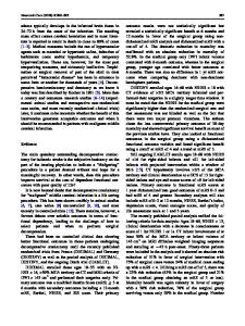
Dear Sir: Recent advances in devices, techniques, and periprocedural patient management in mechanical thrombectomy enable neuro-interventionists to recanalize occluded cerebral arteries with greater efficiency and safety. Image-guided recanalization could extend the therapeutic time window up to 24 h following ischemic stroke [1]. It is conceivable that a subset of patients may benefit from late recanalization, if viable brain tissue remains beyond 24 h [2]. We report a patient who underwent mechanical thrombectomy 96 h after symptom onset with diffusion/perfusion image evaluation. A 56-year-old man with no previous medical history was admitted to the local neurological clinic complaining of a sudden onset left-side weakness. Initial brain magnetic resonance imaging (MRI) revealed acute infarction involving right prefrontal cortex with right middle cerebral artery (MCA) occlusion (Fig. 1a–c). The regular dose of recombinant tissue plasminogen activator was administered within 3 h after symptom onset, but mechanical thrombectomy was not considered
* Jun Soo Byun [email protected] 1
Department of Neurology, Chung-Ang University Hospital, Seoul, Korea
2
Department of Radiology, Chung-Ang University Hospital, Seoul, Korea
because of the mild neurological deficit. The patient was treated with aspirin, clopidogrel, and 80 mg of atorvastatin 24 h after thrombolysis. However, weakness was aggravated after 4 days and neurological examination revealed aggravation of National Institute of Health Stroke Scale from 2 (1 from upper extremity and 1 from lower extremity at initial examination) to 7 (2 from upper extremity, 2 from lower extremity, 1 from dysarthria, 1 from sensory, and 1 from facial palsy at 4th day). The patient was transferred to a tertiary hospital, and followed MRI showed increased extent of acute infarction encompassing right corona radiata and a significant amount of the ischemic penumbra in the right MCA territor
Data Loading...