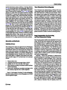
Improving congenital heart disease imaging using 3d whole-heart dual-phase MRI
- PDF / 1,361,735 Bytes
- 2 Pages / 595.276 x 793.701 pts Page_size
- 48 Downloads / 318 Views
POSTER PRESENTATION
Open Access
Improving congenital heart disease imaging using 3d whole-heart dual-phase MRI Tarique Hussain1*, Dirk Lossnitzer1, Sergio Uribe2, Hannah Bellsham-Revell1, Isra Valverde1, Reza Razavi1, Phillip Beerbaum1, Aaron Bell1, Rene Botnar1, Tobias Schaeffter1, Gerald Greil1 From 2011 SCMR/Euro CMR Joint Scientific Sessions Nice, France. 3-6 February 2011 Introduction A single-phase 3d steady-state-free precession whole heart approach with respiratory navigator gating and ECG triggering (3d SSFP) is now frequently used for diagnostic imaging in children with congenital heart disease (CHD). However, certain cardiac structures may be better assessed during the systolic and others in the diastolic rest period. Therefore, a previously described dual phase 3d-SSFP whole heart sequence may offer clinical benefit in these patients. (Uribe et al 2008) Purpose To determine whether 3d-dual phase imaging for CHD offers benefits in demonstrating or measuring cardiovascular structures. Methods 50 consecutive children with CHD underwent 3D SSFP dual-phase imaging. All cardiac chambers and great vessels were analyzed for contrast-to-noise ratio (CNR) and image quality (IQ) (consensus reading by two independent observers; grade 0=non diagnostic; 1 to 4=diagnostic, McConnell et al 1997). Twelve patients were referred for RVOT assessment. Dimensions were measured at valvar, supravalvar and pre-bifurcation levels. CNR, image quality and RVOT measurements were compared between systole and diastole.
The mean HR was 90 bpm (56 to 139). Dual phase yielded diagnostic imaging for all chambers and great vessels in 48 cases. Systolic imaging alone was diagnostic in 45 cases and diastolic imaging in 47 cases. CNR (paired t-test) and IQ (Wilcoxon Signed Ranks test) were significantly (p
Data Loading...