In Situ Real Time Observation of Chemical Vapor Deposition Using an Environmental Transmission Electron Microscope
- PDF / 6,404,173 Bytes
- 10 Pages / 414.72 x 648 pts Page_size
- 75 Downloads / 520 Views
involving CVD offer several advantages over those using physical vapor deposition.
Highly
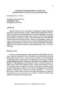
conformal films often result and for several metal systems, selective growth through insulators may allow for reduction of processing steps in multi-level interconnect architectures. As was the case for molecular beam epitaxy several years ago, in situ, real time measurements during growth are increasing our understanding of processes leading to CVD at the atomic and molecular level. It is partially due to the complications of the gaseous and often toxic, rather than UHV, ambient that these in situ, real time studies have only recently been undertaken for CVD. Here, we describe a technique for observing film growth by CVD using processing conditions (pressure, temperature) common for the material system being investigated. A transmission electron microscope (TEM) has been equipped with a differentially pumped environmental cell (E-Cell) which allows the flow of gases through the specimen region during observation. Several metal- insulator systems have been investigated. Here we focus on Al CVD from trimethyl amine alane (TMAA) and Au deposition from ethyl (trimethylphosphine) gold (I), both onto Si0 2 . In the case of Au CVD, observation of beam enhanced growth suggests novel processing techniques involving electron stimulated CVD. EXPERIMENT A Philips 400T TEM operating at 120 keV has been outfitted with a special differentially pumped E-Cell which allows reaction of gases with the sample during imaging. This microscope and E-Cell are described in detail elsewhere [I ] and schematically depicted in Fig. 1. The E-Cell is situated between the upper and lower objective lens pole-pieces and is isloated from the rest of the microscope column by o-rings. Any gases escaping through the 20O01m diameter differential pumping apertures are evacuated by a turbomolecular pump (TMP). Precursors flow from the gas bottle through a shut off and leak valve to the sample space of the microscope. Pressures are 75 Mat. Res. Soc. Symp. Proc. Vol. 404 ©1996 Materials Research Society
Fig. 1. Schematic view of the E-cell. Gases flow from the gas bottle through shut-off and leak
valves which regulate P
TO
G
pressure in the sample space. A thermocouple gauge (TC) measures pressure. The sample
lvI
(S) sits in the polepiece
A(P)
and is isolated from
the microscope vacuum 200 g.m diameter
Sby
differential
L i
pumping
apertures. The sample space is evacuated when the valve to the turbomolecular pump (TMP) is open. This valve is closed during growth.
sensed by thermocouple and cold-cathode gauges. When the valve leading to the TMP is open, the sample space is evacuated. Closing this valve allows flow of gases over the sample during imaging. The base pressure of the E-Cell is the microscope vacuum of 10-7 Torr and high spatial resolution images have been acquired at pressures of several Torr. Use of a standard heating specimen holder allows sample temperatures to reach 1000°C during imaging. Temperatures are measured with an accuracy of ±
Data Loading...