In Situ Testing Using Synchrotron Radiation Computed Tomography in Materials Research
- PDF / 1,342,949 Bytes
- 11 Pages / 432 x 648 pts Page_size
- 65 Downloads / 316 Views
MRS Advances © 2019 Materials Research Society DOI: 10.1557/adv.2019.390
In Situ Testing Using Synchrotron Radiation Computed Tomography in Materials Research Xinchen Ni1, Nathan K. Fritz2, Brian L. Wardle2 1 Department of Mechanical Engineering, Massachusetts Institute of Technology, Cambridge, MA 02139, U.S.A.
2
Department of Aeronautics and Astronautics, Massachusetts Institute of Technology, Cambridge, MA 02139, U.S.A.
ABSTRACT
High resolution (< 1 µm) computed tomography is an attractive tool in materials research due to its ability to non-destructively visualize the three-dimensional internal microstructures of the material. Recently, this technique has been further empowered by adding a fourth (temporal) dimension to study the time-lapse material response under load. Such studies are referred to as four-dimensional or in situ testing. In this snapshot review, we highlight three representative examples of in situ testing using synchrotron radiation computed tomography (SRCT) for composites failure analysis, measurement of local corrosion rate in alloys, and visualization and quantification of electrochemical reactions in lithium-ion batteries, as well as forward-looking integration of machine learning with in situ CT. Lastly, the future opportunities and challenges of in situ SRCT testing are discussed.
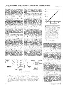
INTRODUCTION In the last few decades, high resolution (15 hours per scan) to achieve sufficient resolution (micrometer or submicrometer scale) for many materials, oftentimes excluding the possibility of in situ tests. On the other hand, the extremely high photon flux of SRCT enables the same level of resolution with a much shorter scan time (typically on the order of minutes per scan), opening up the potential for in situ tests. However, one of the main disadvantages of SRCT is that the beamline is usually a shared facility at a national or supra-national level, with access to the beam time being highly competitive and therefore constrained. In our experience at all 3 Tier 1 beamlines, the average allotted beam time is only 2 – 3 days per visit, and oftentimes only once per year, leaving little room for learning and iteration. Therefore, these two X-ray CT tools are not mutually exclusive and in fact are complementary to each other. See Figure 1 for a high-level comparison between the two. This snapshot review focuses on the use of SRCT to conduct in situ tests. It is worthwhile to note that despite the relatively fast scanning time of SRCT compared to lab-based CT, scanning time remains an outstanding key challenge. First, there are processes, e.g., high strain-rate loading, in which the microstructure change of the material occurs much faster than the imaging rate. In this case, in situ SRCT is unable to capture the important morphology change information. Second, in situ SRCT is a very data-intensive technique, which generates very large datasets (on the order of terabytes) in a short period of time (24 hours), and it takes considerable time to analyse the data post-acquisition. In our experience, 2 –
Data Loading...