Joint line reestablishment in revision total knee arthroplasty
- PDF / 1,625,150 Bytes
- 9 Pages / 595.276 x 790.866 pts Page_size
- 3 Downloads / 339 Views
Arthroplasty
RESEARCH
Open Access
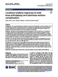
Joint line reestablishment in revision total knee arthroplasty Mustafa Çınar Akça1, Yavuz Akalın1 , Nazan Çevik1 , İsmail Gökhan Şahin2 , Özgür Avcı1 and Alpaslan Öztürk1* Abstract Background: In this study, the traditional “Anatomical Landmark-Distance Method (AL-DM)” in the formation of joint line (JL) was compared with “Adductor Tubercle-Ratios method” (AT-RM), and the effect of reestablishment of JL on clinical and functional outcomes were evaluated. Materials and methods: 16 revision total knee arthroplasties (rTKAs) were performed by using “AT-RM” (group 1) and 16 rTKA by using “AL-DM” (group 2) in our clinic between 2015 and 2018. The data were prospectively collected and a total of 32 knees of 31 patients were analyzed. At the final follow-up, knee functions were evaluated by using Knee Society Score (KSS) knee and function, Western Ontario and McMaster Universities Arthritis Index (WOMAC) scores, Short Form-36 (SF-36) questionnaires and physical examinations. Results: Postoperative flexion arc was higher in Group 1. KSS knee and function scores were better in group 1. In group1, JL was reestablished successfully in all revision rTKAs in terms of ATJL and the tibial tubercle TT-JL ratios. The improvement in KSS knee and function scores and WOMAC scores were also better in group 1. Measurements showed that the improvement in KSS scores increased as AT-JL and TT-JL distances approached the calculated values. Conclusion: “AT-RM” was shown to be superior to the traditional distance method in terms of JL reestablishment. Functional results and patient satisfaction increased when JL was reestablished. Keywords: rTKA, Adductor tubercle, Tibial tubercle, Ratios method, Anatomical landmark, Distance method, Joint line (JL)
Introduction Joint line (JL) restoration is a prerequisite for a successful revision total knee arthroplasty (rTKA) [1]. It has been reported that there was a19 millimeter (mm) JL elevation and a 10 mm depression, even with primary TKA [2]. JL elevation of more than 8 mm was reportedly associated with unfavorable clinical results [3]. Moreover, a recent study showed that elevation over 4 mm was related to lower patellofemoral function [4]. Anatomical structures around the knee, such as medial and lateral epicondyles (ME, LE), tibial tubercle (TT) and fibular head (FH) have been used for calculation * Correspondence: [email protected]; [email protected] 1 Research and Training Hospital Clinic of Orthopaedics and Traumatology, Health Sciences University Bursa YuksekIhtisas, 16310, Yildirim, Bursa, Turkey Full list of author information is available at the end of the article
and restoration of JL in rTKA [5–7]. However, precept distances like ‘2 cm proximal to the fibular head’ or ‘1.5 – 2 cm distal to the lateral epicondyle’ cause JL to be elevated by more than 5 and 8 mm in rTKA because there are obvious variations in terms of race, gender and body mass index [8–14]. Given all these disadvantages, some new techniques, such as methods of ratios of f
Data Loading...