Left upper lung cancer with persistent left superior vena cava and left azygos vein: a case report
- PDF / 3,466,003 Bytes
- 4 Pages / 595.276 x 790.866 pts Page_size
- 23 Downloads / 289 Views
(2020) 15:254
CASE REPORT
Open Access
Left upper lung cancer with persistent left superior vena cava and left azygos vein: a case report Zhongben Tang1* , Yin Teng1, Jian Li1, Xiaojun Du1, Jiarong Xiao1 and Gongshun Tang2
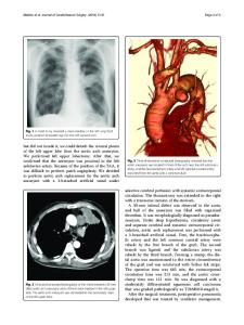
Abstract Background: With the popularization of thoracoscopic surgery, more and more macrovascular malformations have been reported. Understanding some vascular malformations with relatively fixed anatomical site and their range of drainage could avoid severe complications during the surgery. Persistent left superior vena cava (PLSVC) is a common thoracic vascular malformation, and is always combined with other cardiovascular dysplasia. As for the patient with upper left lung cancer in this case, he had PLSVC and left azygos vein, and non-metastatic enlargement of the lymph nodes at the same time, which had influenced the decisions on surgery and treatment. We made a summary of experience regarding this. Case presentation: A 46-years-old male patient, his CT found a space-occupying lesion in the superior lobe of the left lung. The chest CT showed that the patient had PLSVC and left azygos vein, and multiple enlarged lymph nodes in the mediastinum. The patient received thoracoscopic upper left lung lobectomy and lymph node dissection. It was discovered that the left azygos vein had a concealed form, which influenced the lymph node dissection. The postsurgery pathology showed that there was squamous cell carcinoma in the upper left lung (pT2bN0M0 p Phase IIA) and no cancer metastasis with the lymph nodes. The patient had a good post-surgery recovery. Conclusions: PLSVC is not rare, and is always combined with other vascular malformations. If discovering PLSVC before surgery, we suggest completing chest enhanced CT and vascular reconstruction, to find out other cardiovascular malformations that may exist. Left azygos vein is a rare vascular malformation, but it has a relatively fixed anatomical site, and always co-exists with PLSVC, therefore, understanding anatomy of left azygos vein is good for preventing accidental damage. Especially when performing surgery above the left pulmonary artery trunk, attention shall be paid to preventing damage to the left azygos vein. In addition, as for the patient with the diagnosis of lung cancer before surgery, it is not reliable to judge whether there is metastasis or not merely according to the size of the lymph nodes, instead, PET-CT or needle biopsy is recommended. Keywords: Lung cancer, Persistent left superior vena cava, Left azygos vein, Surgery
* Correspondence: [email protected] 1 Department of Thoracic, The Affiliated Hospital of Guizhou Medical University, 28 Guiyi Street, Guiyang 550004, Guizhou, China Full list of author information is available at the end of the article © The Author(s). 2020 Open Access This article is licensed under a Creative Commons Attribution 4.0 International License, which permits use, sharing, adaptation, distribution and reproduction in any medium or format, as long as you give appropriate credit to the original author(s
Data Loading...