Magnetic Spectro-Microscopy Using Magneto-Dichroic Effects in Photon-Induced Auger Electron Emission
- PDF / 1,003,534 Bytes
- 6 Pages / 420.48 x 639 pts Page_size
- 14 Downloads / 331 Views
MAGNETIC SPECTRO-MICROSCOPY USING MAGNETO-DICHROIC EFFECTS IN PHOTON-INDUCED AUGER ELECTRON EMISSION
3 2 2 3 4 C.M. SCHNEIDER1. , K. MEINEL , K. HOLLDACK , H.P. OEPEN , M. GRUNZE , 2 AND J. KIRSCHNER I Surface Physics Laboratory, Simon Fraser University, Burnaby, B.C., V5A IS6, Canada 2 MPI f. Mikrostrukturphysik, Weinberg 2, 0-4050 Halle/Saale, Germany 3 Inst. f. Angew. Phys. Chemie, Universitdit Heidelberg, Im Neuenheimer Feld 253, D-6900 Heidelberg, Germany 4 Inst. f. Grenzflachenforschung u. Vakuumphysik, KFA Jilich, Postfach 1915, D-5170 Jtilich, Germany
ABSTRACT We have imaged the magnetic domain structure on the surface of Fe(100) single crystals using energy resolved photoemission microscopy with circularly polarized soft x-rays. The contrast between different domains arises due to magneto-dichroic effects in the emitted Auger electrons. This new approach offers a surface sensitive way to combine chemical and magnetic information on a microscopic scale.
INTRODUCTION Within the last decades photoelectron spectroscopy has matured into a powerful tool in surface science. Even the photoelectron spin has been successfully made accessible to experimental investigations on both non-magnetic and ferromagnetic materials [I]. Spin-split electronic states in ferromagnets may be studied in great detail either by an explicit analysis of the photoelectron spin [2], or by means of the recently reported magnetic circular dichroism in photoemission (MCDAD) [3]. Both approaches add magnetic sensitivity to conventional photoelectron spectroscopy. The present photoemission experiments are usually hampered by sampling a rather 2 large surface area (-mm ), as determined by the size of the illuminated spot and the characteristics of the electron optics. The investigation of electronic and magnetic microstructures, however, requires lateral resolution capabilities. The combination of lateral resolution and magnetic sensitivity in photoemission techniques can be used in two ways: magnetic sensitive spectroscopy on well-defined areas on the surface (micro-spectroscopy), and imaging of magnetic domains with electrons of a selected kinetic energy (spectro-microscopy).
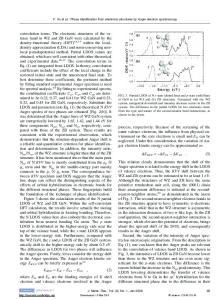
EXPERIMENTAL DETAILS Our experimental approach to the problem employed a so-called ESCASCOPE, which has been described in detail by Coxon et al. [4]. Briefly, the set-up features a hemispherical energy analyzer (HMA) in combination with an electrostatic input lens system. The instrument can be operated either in a spectroscopic mode as a conventional electron spectrometer, or in an imaging mode. The latter allows the acquisition of photoelectron images with a spatial resolution Ad
Data Loading...