Metastatic bronchogenic carcinoma with myocardial and endocardial secondaries
- PDF / 775,855 Bytes
- 3 Pages / 595.276 x 790.866 pts Page_size
- 35 Downloads / 249 Views
CASE IMAGE IN CARDIOVASCULAR ULTRASOUND
Metastatic bronchogenic carcinoma with myocardial and endocardial secondaries Jomy Vadasseril Jose1 · Gopalan Nair Rajesh1 Received: 31 August 2020 / Revised: 28 September 2020 / Accepted: 10 October 2020 © Japanese Society of Echocardiography 2020
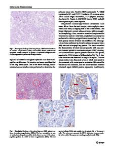
A 68-year-old male, an exsmoker and hypertensive, presented to us with chest pain not typical of angina since the past 3 days. He was on palliative chemotherapy and radiotherapy for stage IV bronchogenic carcinoma (squamous cell carcinoma) for the past 3 months. His ECG showed significant ST elevation in precordial leads suggestive of acute anterior wall myocardial infarction. His troponin I was elevated. As shown in Fig. 1, transthoracic echocardiography revealed a necrosed, echolucent focus in the anterolateral wall of the left ventricle (LV). There was no evidence of communication with the LV cavity. There was also another echogenic pedunculated mass protruding into the LV from the anterolateral wall. A CT scan was carried out which revealed that the myocardium of the apex and lateral wall of the LV was replaced by heterogenous, hypodense peripherally enhancing mass lesion suggestive of metastatic deposits. There was also minimal loculated pericardial effusion. The primary lesion was noted in the anterior segment of the left upper lobe of the lung close to the mediastinum along with bilateral pleural effusion (see also Movie 1, 2, 3). Patient was already having multiple metastases and his prognosis was very poor. He was also not willing for further investigations. He was treated with antiplatelets, statins, analgesics, and supportive care. He had sudden cardiac death at his home, 23 days from the date of echocardiography.
Autopsy studies show that bronchogenic carcinoma is the primary tumor in 36% of patients with cardiac metastases [1]. Bronchogenic carcinoma may involve the heart and pericardium by direct extension or by a combination of lymphatic and hematogenous dissemination [2]. Endocardial metastases from lung carcinoma as in this case are very rare [3]. Mechanism of invasion is through the bloodstream into the heart’s chambers with intracavitary lodging. Endocardial metastasis secondary to diffusion from myocardial metastases is also possible [3]. Mechanisms described for the ST-T changes in cardiac metastases are compression of the coronary arteries with resulting ischemia, a direct extension into the lumen of the coronary arteries or coronary embolization by tumor fragments, neoplastic invasion of the pericardium resulting in a subacute pericarditis and continuous myocardial injury by direct pressure [4]. This case depicts a rare cause of ST elevation in the ECG and highlights the significance of multimodality imaging before deciding on management of the same. Possible mechanisms of sudden cardiac death in this patient could be ventricular free wall rupture, ventricular arrhythmias, or embolization of the pedunculated mass.
Electronic supplementary material The online version of this article (https://doi.org/10
Data Loading...