Microstructural Characterization of Nanosized Ceria Powders by X-Ray Diffraction Analysis
- PDF / 989,622 Bytes
- 9 Pages / 593.972 x 792 pts Page_size
- 2 Downloads / 311 Views
WADAYS different routes such as mechanical alloying, combustion synthesis, plasma forming, explosive forming, electrodeposition, and sol-gel process are used for producing nanosized ceramics or metals. Among them, high energy ball milling (HEBM) is the most useful technique for producing nanosized ceramics and ceramic composites. The ball milling technique is environmentally safer than the method of chemical synthesis, producing far less chemical waste.[1] A number of nanostructured metal oxides and their solid solutions such as Fe2O3-SnO2,[2] ZrO2-Fe2O3,[3] TiO2Fe2O3,[4] TiO2-WO3,[5] Pb(Zr0.52Ti0.48)O3,[6] SrTiO3,[7] SiC, Si3N4, and quartz (SiO2) powders[8] were prepared by using the HEBM method. The crystallite size of the ceramic powders was analyzed mainly by the Scherrer or modified Scherrer formula.[9–12] It was reported in the literature that incorporation of nanosized ceramic particles such as ceria in a metallic matrix, such as Ni, can significantly enhance the hardness, thermal stability, corrosion resistance, and wear resistance of Ni matrix.[13,14] The objective of the present investigation is to apply different X-ray diffraction (XRD) analyses, i.e., Rietveld, classical, and modified Williamson–Hall method, to determine the microstructural parameters of the ball-milled nanosized ceria powders.
II.
EXPERIMENTAL PROCEDURES
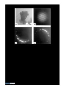
A. Synthesis of Ceria Powders by HEBM Technique HEBM of ceria powder (Alfa Aesar, 99.5 pct) is carried out using cemented tungsten carbide milling media with toluene as the process control agent. The mill is operated at a speed of 300 rpm and the ball-topowder ratio is 10:1. The 0, 1, 3, 5, and 10 hour ballmilled powder is washed with distilled water and then with ethyl alcohol followed by drying. B. Materials Characterization All the XRD experiments are carried out in an XRD machine (Bruker axe D8, Ducom Instrument Pvt. Ltd. AG, Bangalore, India), using 40 kV voltage, 30 mA current, 0.2 mm receiving slit, scintillation counter detector, and Co Ka radiation. But no monochromator is attached with this system. The powders were examined using a field emission scanning electron microscope (Zeiss, Carl Zeiss SMT AG, Oberkochen, Germany) operating at 5.0 kV attached with an energy dispersive X-ray spectrometer (EDS). A high resolution transmission electron microscope (JEOL* 2010F) operating at 200 KV is used to find the *JEOL is a trademark of Japan Electron Optics Ltd., Tokyo.
RANJAN SEN, Research Scholar, SIDDHARTHA DAS, and KARABI DAS, Professors, are with the Department of Metallurgical and Materials Engineering, Indian Institute of Technology, Kharagpur-721302, India. Contact e-mail: [email protected]. ernet.in Manuscript submitted April 13, 2010. Article published online December 1, 2010 METALLURGICAL AND MATERIALS TRANSACTIONS A
crystallite size of the ceria powder. The ceria powder is dispersed in acetone and the solution is kept in an ultrasonic vibrator for 1 hour. The solution is then allowed to settle down for 2 minutes, and a drop of the solution is taken from the top layer and dro
Data Loading...