Microstructural investigation of the oxidation of an Fe-3 pct Cr alloy
- PDF / 4,848,357 Bytes
- 10 Pages / 594 x 774 pts Page_size
- 1 Downloads / 310 Views
I.
INTRODUCTION
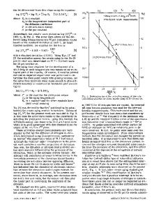
O X I D A T I O N resistant Fe-base alloys generally contain at least one element, e.g., Cr, A1, Si, which is selectively oxidized to form a stable protective oxide on the alloy surface. Oxidation of pure Fe above 600 ~ produces a three layer scale of Fel.xO/Fe304/Fe203. In Fe-Cr alloys the formation of the Fe~_xO wustite layer is suppressed. Depending on the Cr-concentration, the Fe3Oa magnetite layer may be replaced by a spinel, Fe(Fe2.xCrx)O4, and the Fe203 hematite may be replaced by the sesquioxides, (Fe,Cr)203 and Cr203. It is well established that Cr, alloyed in sufficient amounts, imparts good oxidation resistance. ~-s Low Cr concentrations, however, result in poor oxidation resistance, internal oxidation, and nonreproducible weight-gain vs time measurements. 9 The mechanisms responsible for scale development are not well understood, and numerous theories have been postulated. 6J~ The types of oxides formed are also controversial. An investigation has therefore been designed to study the Fe-Cr system by high resolution analytical electron microscopy techniques. The purpose of the research was to investigate the oxide nucleation mechanism, the types of oxides formed, and the growth of these oxides, and to compare these results with the oxidation mechanism of pure Fe. In the present paper we will focus on the Fe-3 wt pct Cr system. The oxide scale which develops on this alloy results in poor oxidation resistance and is different from the scales which form on the higher Cr alloys.
carried out so that all specimens had an identical air-formed oxide film on the polished surface prior to the high temperature oxidation experiment. 3 mm discs were punched from the strips; some were jet polished to perforation with 95 pct glacial acetic acid and 5 pct perchloric acid for in-situ oxidation experiments in a Hitachi HU-650B high voltage electron microscope while others were left as 'bulk' specimens to be oxidized first (ex-situ) and then back-thinned to examine the oxide scale. The strips and discs were oxidized under the conditions given in Table I. Flowing, dry oxygen was used for ex-situ oxidations at 3 kPa pressure. TEM column vacuum (--~1 mPa) was the environment for the oxidations on thin TEM foils. Back-thinning was performed by protecting one side of the oxidized disc with clear plastic, mounting the disc in a twin-jet polishing unit or in an ionthinner with only one gun operating, and thinning until perforation occurred. In this way it was possible to examine the oxide scale in parallel sections and, if thin enough, to examine the metal/oxide interface. Some of the strips were mounted in epoxy to be cut and polished for transverse sections; others were examined in a Cambridge Stereoscan scanning electron microscope (SEM) and in a PHI 545 scanning Auger system. The oxidized 3 mm discs were examined in a Philips EM400T transmission electron microscope (TEM). Energy dispersive X-ray analysis on an EDAX 60 system was performed in the SEM and TEM. Standard ZAF quantitative analysis was Table I.
Data Loading...