Microstructure and Characteristics in the Organic Matrix Layers of Nacre
- PDF / 146,397 Bytes
- 4 Pages / 612 x 792 pts (letter) Page_size
- 31 Downloads / 316 Views
The direct observation of a type of microstructure in the organic matrix layers of nacre was obtained with a transmission electron microscope. The microstructure, which is referred to as mineral bridge in the biomineralization, is nanoscale and randomly distributed in the layers. Statistical analysis gives the distribution laws and characteristics of mineral bridges in the organic matrix layers. The existence of mineral bridges in nacre was confirmed, and it was shown that the microarchitecture of nacre should be described as a “brick–bridge–mortar” arrangement rather than traditional “brick and mortar” one.
Nacre, one of several kinds of molluscan hard tissue, is considered as a ceramic composite containing 95 vol% interlocking aragonite platelets staggered in successive laminae and separated by a 5% organic matrix. Since it can give a conceptual guidance to the biomimetic design of synthetic materials, a great deal of attention has been attracted to the microstructure of nacre in recent years.1– 4 The traditional model of nacre is considered as a “brick and mortar” (BM) arrangement. It is the unique arrangement that is believed to result in lightweight materials with high mechanical performance.5–8 However, the study of the microstructure of nacre has gained some significant developments in recent years. In particular, Schaffer et al.9 clearly observed many nanopores in the interlamellar organic matrix sheets of nacre in terms of various microscopic observations and then gave a statistical distribution. Consequently, they supported the
model of nacre growth that is based on mineral bridges between successive aragonite platelets. Furthermore, according to their transmission electron microscopy (TEM) micrograph, they stated that “these (gray in the micrograph) may be mineral bridges between the nacre plates, but it is difficult to be certain of this assignment.” Here we describe direct observation of mineral bridges in the organic matrix layers of nacre. And by using statistical analysis, we obtain the characteristics and the distribution law of the microstructure. It is interesting that results presented in this paper are consistent with an earlier estimate.9 To reveal the microstructure of nacre, all observations of nacre were performed with an H-8100 TEM at an accelerating voltage of 200 kV. The testing samples are the nacre of Haliotis iris shell, an abalone shell from New Zealand. The keratin layer of the shell was
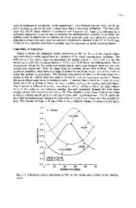
FIG. 1. (a) TEM image showing the microarchitecture of the cross sectional surface of nacre. It appears to be a traditional “brick and mortar” arrangement. (b) [boxed area in (a)] TEM image of the organic and inorganic layers on the cross sectional surface of nacre. There exist some mineral bridges in the organic matrix layers. The positions of the mineral bridges in the layers are random. (c) [boxed area in (b)] TEM image of a mineral bridge between two successive platelets of nacre. The appearance of mineral bridges approximates circular column. J. Mater. Res., Vol. 17, No. 7, Jul 2002
http://jo
Data Loading...