Neuro-Sweet syndrome - a rare differential diagnosis in aseptic meningoencephalitis
- PDF / 728,993 Bytes
- 3 Pages / 595.276 x 790.866 pts Page_size
- 87 Downloads / 298 Views
(2019) 1:36
LETTER TO THE EDITOR
Neurological Research and Practice
Open Access
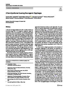
Neuro-Sweet syndrome - a rare differential diagnosis in aseptic meningoencephalitis Elgin Hoffmann1,2*, Christian Boßelmann1, Stephan Forchhammer3, Holger Lerche1 and Tobias Freilinger1,4 Abstract Acute febrile neutrophilic dermatosis (Sweet‘s syndrome) is a dermatological entity, which may be associated with malignancies, drugs, and infections and which is characterized by high fever, elevated neutrophils, and tender erythematous skin lesions. Involvement of the nervous system – Neuro-Sweet syndrome (NSS) - is rare, manifesting most commonly with an encephalitic syndrome in addition to fever and dermal lesions. Here, we report an unusual case of NSS in a Caucasian male patient in the setting of B-cell-lymphocytosis, with encephalitis preceding dermal lesions. Symptoms resolved completely in response to corticoids. NSS is a rare, but important differential diagnosis in the work-up of febrile aseptic meningoencephalitis unresponsive to anti-infectious treatment. Due to its rarity and clinical variability, diagnosis of NSS might be challenging. Knowledge of this entity may facilitate proper diagnosis and differentiation from conditions with similar clinical presentation, especially Neuro-Behçet‘s disease. It may further lead to early detection of a potentially underlying malignancy and help in initiating adequate therapy. Keywords: Differential diagnosis to meningoencephalitis, Neuro-sweet syndrome, Neuro-Behçet disease, Paraneoplastic syndromes Dear Editors, We would like to report a case of Neuro-Sweet syndrome (NSS) as a rare but important differential diagnosis of aseptic meningoencephalitis in the setting of B-cell-lymphocytosis, with encephalitis preceding dermal lesions. A previously healthy 75-year-old male developed psychomotor retardation, altered state of consciousness, and fatigue for 2 weeks. There were no focal deficits, but high fever (> 39 °C) with isolated CRP-elevation (13.7 mg/dl). Erythematous plaques on both thumbs had evolved 2 days prior to presentation at the hospital (Fig. 1a). There were no oral, genital or ophthalmological lesions. cMRI was unremarkable except for leukencephalopathy. CSF analysis showed mild pleocytosis (8/μl). Microbiological/serological analysis of CSF and blood was unremarkable. Whole-body CT revealed splenomegaly and pronounced medullary cavities. Fever and CRP* Correspondence: [email protected] 1 Department of Epileptology, University Hospital for Neurology, Eberhard Karls University, Hoppe-Seyler-Str. 3, 72076 Tübingen, Germany 2 Current address: University Hospital for Radiation Oncology, Eberhard Karls University, Hoppe-Seyler-Str. 3, 72076 Tübingen, Germany Full list of author information is available at the end of the article
elevation persisted despite calculated anti-infectious therapy, with increasing neutrophilic count (leukocytes: 8720/μl, neutrophils: 6120 /μl, 70,1% neutrophils) and dermal lesions spreading to other areas. Skin biopsy revealed neutrophilic
Data Loading...