Optimized Silver Film over Nanosphere Surfaces for the Biowarfare Agent Detection Based on Surface-Enhanced Raman Spectr
- PDF / 471,413 Bytes
- 6 Pages / 612 x 792 pts (letter) Page_size
- 48 Downloads / 265 Views
R8.54.1
Optimized Silver Film over Nanosphere Surfaces for the Biowarfare Agent Detection Based on Surface-Enhanced Raman Spectroscopy Xiaoyu Zhang and Richard P. Van Duyne Department of Chemistry, Northwestern University, Evanston, IL 60208-3113, USA ABSTRACT This work presents the rapid detection of Bacillus subtilis spores, harmless simulants for Bacillus anthracis, using surface-enhanced Raman spectroscopy (SERS) on silver film over nanosphere (AgFON) substrates. Calcium dipicolinate (CaDPA), a biomarker for bacillus spores, can be extracted effectively from spores with nitric acid and successfully detected by SERS. The highly tunable nature of AgFON optical properties was exploited to establish general optimization conditions. AgFON surfaces optimized for 750-nm laser excitation have been characterized by UV-vis diffuse reflectance spectroscopy. The SERS signal from extracted CaDPA was evaluated over the spore concentration range 10-15-10-12 M to determine the adsorption capacity of the AgFON surface and the limit of detection (LOD). These sensing capabilities have been successfully transitioned to an inexpensive, portable Raman spectrometer. Using the extraction method and this field-portable instrument, the anthrax infectious dose of 104 spores were detected with only a 5-second collection period on a one-month-old prefabricated AgFON substrate. INTRODUCTION Vibrational spectroscopic methods are valuable analytical tools because they yield not only quantitative information but also unique vibrational signatures for small molecule analytes. Raman spectroscopy, in all its forms, is a vibrational spectroscopic method that has the inherent ability to distinguish between molecules with great similarity. Unfortunately, high laser powers and long acquisition times are usually required to achieve high quality Raman spectra due to the
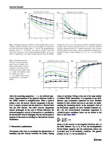
Figure 1. Nanosphere lithographic fabrication of nanoparticle arrays and film over nanosphere surfaces (FON).
R8.54.2
inherently small normal Raman scattering (NRS) cross section of many molecules of interest.[1] Higher intensity Raman signals and lower detection limits can be achieved using SERS. SERS produces very large enhancements in the effective Raman cross section of species spatially confined within zone of the electromagnetic fields (viz. 0–4 nm)[2] generated upon excitation of the localized surface plasmon resonance (LSPR) of nanostructured noble metal surfaces. This large electromagnetic field induces a dipole in nearby molecules, thus enhancing Raman scattering from absorbed molecules. The Raman signals of ensemble-averaged molecules show enhancement of up to 8 orders of magnitude[3], while the signals from single molecules can show an increase by 14 to 15 orders of magnitude in special cases[4, 5]. In comparison with infrared and NRS spectroscopies, SERS enjoys the advantages of application in aqueous media and the sensitivity sufficient for trace level detection[6]. For sensors, it is important that the optical properties of the substrate be designed to fully maximize
Data Loading...