Painful left forearm swelling
- PDF / 170,868 Bytes
- 2 Pages / 595.276 x 790.866 pts Page_size
- 95 Downloads / 434 Views
TEST YOURSELF: ANSWER
Painful left forearm swelling Julio Brandão Guimaraes 1,2,3 Henrique Lederman 1,2
&
Renato Masson 2 & Marcelo Petrilli 4 & Maria Teresa de Seixas Alves 5 &
# ISS 2020
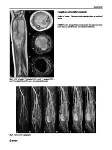
Final diagnosis: Subperiosteal Hemophilic Pseudotumor Discussion Anteroposterior and lateral radiographs of the left forearm demonstrate (Fig. 1) expansile subperiosteal lytic bone lesions that extend into the soft tissue, associated with diffuse cortical thinning, subperiosteal scalloped erosion of the radius, and radiating trabeculae with periosteal reaction. Imaging findings also indicate the presence of calcifications with a curvilinear pattern in the periphery of soft tissues in the medial proximal radius and bone remodeling in the distal ulna. MRI findings (Fig. 2) show a large encapsulated subperiosteal soft tissue mass surrounding the radius, with distinct signal intensities on both T1- and T2-weighted images, including spontaneous high signal intensity on T1, suggesting different stages of hemorrhage. Furthermore, the interosseous space is widened due to the dimension of the soft tissue mass. Post-contrast imaging did not reveal any solid The case presentation can be found at doi: 10.1007/s00256-020-03590-x * Julio Brandão Guimaraes [email protected]; [email protected] 1
Department of Radiology, Grupo de Apoio ao Adolescente e à Criança com Câncer (GRAACC), Sao Paulo, Brazil
2
Department of Radiology, Federal University of Sao Paulo (UNIFESP), Sao Paulo, Brazil
3
Musculoskeletal and Quantitative Imaging Research Group (MQIR), Department of Radiology and Biomedical Imaging, University of California, San Francisco, 185 Berry St, Suite 350, San Francisco, CA 94158, USA
4
Department of Orthopedic Surgery, Grupo de Apoio ao Adolescente e à Criança com Câncer (GRAACC), Sao Paulo, Brazil
5
Department of Pathology, Federal University of Sao Paulo (UNIFESP), Sao Paulo, Brazil
areas, exhibiting a predominant peripheral enhancement distribution pattern. MRI angiography findings (Fig. 3) show an active filling of a cystic cavity within the lesion, suggesting an intracystic hemorrhage that had been actively bleeding. The pathology of the biopsy sample confirmed the content of the pseudocyst’s cavities, which were filled with hemorrhagic and fibrinoid content (Fig. 4), as well as negative for atypical cells. Subsequently, the patient underwent surgery with curettage and iliac bone grafting, which was shortened by a massive bleeding of the donor site for the graft (Fig. 5). After a severe drop in hemoglobin, coagulation tests for factor VIII deficiency were carried out, and the patient was diagnosed with type A hemophilia. Pathology findings, along with the newly uncovered history of type A hemophilia, are consistent with the diagnosis of a hemophilic pseudotumor. The patient showed clinical improvement after the replacement of factor VII and management of the anemia, and 6 months into recovery, there were no signs of local recurrence (Fig. 6). Type A hemophilia is an X-linked recessive gene
Data Loading...