Painful type II os naviculare: introduction of a standardized, reproducible classification system
- PDF / 3,161,228 Bytes
- 9 Pages / 595.276 x 790.866 pts Page_size
- 63 Downloads / 327 Views
SCIENTIFIC ARTICLE
Painful type II os naviculare: introduction of a standardized, reproducible classification system Sarah I. Kamel 1 & Jeffrey A. Belair 1 Adam C. Zoga 1
&
Tarek M. Hegazi 2 & Ethan J. Halpern 1 & Vishal Desai 1 & William B. Morrison 1 &
Received: 30 March 2020 / Revised: 1 June 2020 / Accepted: 3 June 2020 # ISS 2020
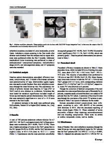
Abstract Objective To provide a novel MRI classification system for the symptomatic type II os naviculare by creating a standardized grading of associated bone marrow edema (BME) and correlating with patient symptoms. Methods BME was classified on an ordinal scale: grade 1, faint signal immediately adjacent to the synchondrosis; grade 2, intermediate signal within the os and navicular tuberosity without extending to the navicular body; grade 3, intense signal extending to the navicular body. BME on 59 MRIs was independently graded by three radiologists. Inter- and intra-observer agreement was analyzed using intraclass correlation coefficient. Univariate and multivariate analyses assessed for patient and imaging characteristics predictive of subjective pain score. A cohort of 82 patients without BME represented a control group. Results Inter-observer agreement of BME grade was 0.95 (CI 0.93–0.97) and intra-observer was 0.92 (CI 0.87–0.96), indicating excellent agreement. In patients with BME, predictors of more severe pain were longer duration of pain (p = 0.02) and presence of soft tissue edema overlying the os naviculare (p < 0.001). One hundred percent of subjects with BME localized their pain to the medial midfoot (59/59) versus 25.6% (21/82) of controls (p < 0.001). Conclusions This novel grading system provides reliable quantification of BME associated with os naviculare, which is a specific cause of medial foot/ankle pain. Early diagnosis is important as pain severity worsens with longer duration of symptoms. Pain severity is correlated with soft tissue edema overlying the os, which may be secondary to extrinsic compression, reactive to biomechanical stress, or reflect direct trauma. Keywords Painful type II os naviculare . Accessory navicular syndrome . Medial foot pain . Synchondrosis . Bone marrow edema . Posterior tibial tendinosis
Introduction The os naviculare (also referred to as the accessory navicular or navicular os), similar to other accessory ossicles within the foot, is a normal anatomic variant and is generally discovered
incidentally. The overall incidence of the accessory navicular has been reported between 4 and 21%, is frequently bilateral and first appears in adolescence, with a higher prevalence in females [1–4]. First described by Emil S. Geist in 1914 and further refined in publications by Sella et al., the accessory
* Jeffrey A. Belair [email protected]
William B. Morrison [email protected]
Sarah I. Kamel [email protected] Tarek M. Hegazi [email protected]
Adam C. Zoga [email protected] 1
Department of Musculoskeletal Radiology, Thomas Jefferson University Hospital, Main Building 1090, Philadelphia, PA
Data Loading...