Pearls on resecting the TMJ chondrosarcoma: how i do it
- PDF / 1,216,008 Bytes
- 5 Pages / 595.276 x 790.866 pts Page_size
- 71 Downloads / 337 Views
HOW I DO IT
Pearls on resecting the TMJ chondrosarcoma: how i do it Zhong Jie Chia1 · Yijin Jereme Gan2 · Ming Yann Lim2 Received: 10 August 2020 / Accepted: 16 September 2020 © Springer-Verlag GmbH Germany, part of Springer Nature 2020
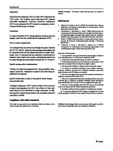
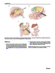
Abstract Background Chondrosarcoma of the head and neck is uncommon and Chondrosarcoma of the Temporomandibular Joint is exceedingly rare. We present a 67-year-old male with a large right TMJ Chondrosarcoma. Methods Patient underwent right segmental mandibulectomy with excision of TMJ tumor, lateral temporal bone resection and right infratemporal fossa resection with Vastus Lateralis flap reconstruction of facial defect. Steps and technical considerations are presented in the video attached. Conclusion This case and video illustration show the technical feasibility of complete resection of Chondrosarcoma of the TMJ while preserving function of critical structures most importantly the ophthalmic branch of the facial nerve. Keywords Chondrosarcoma · Temporomandibular joint · Techinical considerations · Infratemporal fossa
Introduction
Case presentation
Chondrosarcoma is a malignant tumor characterized by the formation of cartilage by tumor cells [1] and accounts for 11% of all primary malignant bone tumors [2]. Chondrosarcoma of the head and neck is an uncommon disease and makes up 0.1% of all head and neck neoplasms [3]. Chondrosarcoma of Temporomandibular Joint is exceedingly rare. Due to the presence of several vital structures around the TMJ, treatment for Chondrosarcoma is a difficult task. A balance must be struck between tumor resection and preservation of critical structures. We present a case of Chondrosarcoma of the TMJ, along with resection of the tumor with technical considerations for optimal resection, which will be discussed later.
A 67-year-old Chinese male was referred to our clinic with right temporomandibular joint mass that was associated with increasing pain, which was present for a year prior to the visit. There was an absence of dysphagia, odynophagia and facial weakness. The patient was a non-smoker, non-drinker and had no family history of head and neck cancer. Upon examination, a firm, hard 2 cm mass over the right temporomandibular joint was palpated. There was an absence of trismus and facial nerve function was intact. Oral cavity and oropharynx were normal. Computed Tomography (CT) Face and Magnetic Resonance Imaging (MRI) Neck with contrast were performed. Both scans revealed an irregular, lobulated mass centered on the right temporomandibular joint, which was approximately 4.0 × 2.9 × 2.5 cm and extended anteromedially into the right masticator space and laterally into the right parotid space. This mass had multiple foci of calcifications which demonstrated “rings” and “arcs” configuration. The case was discussed at a multidisciplinary tumor board and there was consensus that the most likely diagnosis was that of TMJ chondrosarcoma. Other differentials included osteoma, osteosarcoma, chondroblastoma. chondromyxoid fibroma and fibrous dysplasi
Data Loading...