PET/CT in a patient with cardiac paraganglioma
- PDF / 965,934 Bytes
- 5 Pages / 595.276 x 790.866 pts Page_size
- 52 Downloads / 343 Views
CASE-IN-POINT
PET/CT in a patient with cardiac paraganglioma Yanhua Duan1 · Rui Xu2 · Wen Liu3 · Xiao Cui1 · Kun Li1 · Xiaoqing Yang4 · Qinghong Shi1 · Zhaoping Cheng1 · Li Chen5 Received: 3 November 2020 / Accepted: 8 November 2020 © Springer Nature B.V. 2020
Abstract A 22-year-old female with SDHB-positive who presented with palpitation and hypertension after adrenalectomy was performed 18F-FDG PET/CT to detect the primary ectopic pheochromocytoma (PCC) and rule out metastasis. PET/CT is useful for detecting and localizing the primary ectopic PCC. Keywords Cardiac paraganglioma · PET/CT · 18F-FDG Pheochromocytoma (PCC) is a rare neuroendocrine tumor, most of PCCs locate in the adrenal medulla [1]. Paragangliomas (PGLs) are extra-adrenal PCCs that arise from chromaffin cells of the paresympathetic or sympathetic ganglia, only 1–2% of PGLs occur in the chest, mostly in the posterior mediastinum [2]. Cardiac PGL is an extremely rare tumor and forming 2% of PGLs [3]. Most of them are benign, highly vascular catecholamine-secreting tumor. A 22-year-old female presented with paroxysmal palpitation, fatigability and hyperidrosis for 1 year, headache and dizziness for 5-month, and was found to have significant
* Zhaoping Cheng [email protected] * Li Chen [email protected] 1
Department of PET/CT, The First Affiliated Hospital of Shandong First Medical University, Shandong Provincial Qianfoshan Hospital Affiliated To Shandong University, Jinan 250014, People’s Republic of China
2
Department of Cardiology, The First Affiliated Hospital of Shandong First Medical University, Jinan 250014, People’s Republic of China
3
Department of Radiology, The First Affiliated Hospital of Shandong First Medical University, Jinan 250014, People’s Republic of China
4
Department of Pathology, The First Affiliated Hospital of Shandong First Medical University, Jinan 250014, People’s Republic of China
5
Department of Ultrasound, Shandong Provincial Hospital Affiliated to Shandong First Medical University, Shandong University, No. 324 Jingwu Road, Jinan 250021, People’s Republic of China
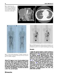
hypertension (180/110 mmHg) for 2-month. Serum and urinary metanephrine and catecholamine levels were significantly elevated (Table 1). PCC was suspected. However, non-contrast MR of abdomen showed a small left adrenal adenoma. The blood pressure increased markedly again after left adrenal gland surgery. Genetic testing was performed and showed a likely pathogenic mutation in the succinate dehydrogenase B (SDHB) gene which associated with PCC and PGL syndromes. 18F-FDG PET/CT was performed to detect the primary ectopic PCC and rule out metastasis. PET/CT (EXPLORER total-body PET/CT scanner) showed a large mass with an diameter of 39 mm × 49 mm × 58 mm on the top of the left atrium (LA) with hyper-metabolism, the maximum standardized uptake value (SUVmax) is about 29.5, and no metastatic lesions were observed (Fig. 1).The higher 18F-FDG-uptake is possibly related to the SDHB mutation [4]. An echocardiogram was performed to de
Data Loading...