Photoluminescence and Photoluminescence Excitation Spectroscopy of In Situ Er-Doped and Er-Implanted GaN Films Grown by
- PDF / 954,370 Bytes
- 7 Pages / 417.6 x 639 pts Page_size
- 47 Downloads / 381 Views
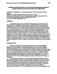
PLE and PL of these two different HVPE-grown samples are compared here first with the Erimplanted MOCVD-grown sample and then the differences and similarities among the siteselective PLE and PL spectra of these three different Er-doped GaN films are discussed in detail. EXPERIMENTAL PROCEDURE The GaN films were doped in-situ with Er in a horizontal HVPE reactor during growth. A peak Er concentration of 2 x 10¹⁹ ions/cm 3 was achieved in this in situ Er-doped GaN at a thickness of 1000 nm [7]. The GaN films grown on sapphire by HVPE were implanted with a dosage of 2 x 10¹⁴ ions/cm2 at 300 keV. The peak concentration of Er is 5.3 x 10¹⁹ ions/cm3 at a depth of 33 nm [7]. These Er-implanted HVPE-grown GaN films were annealed in a conventional tube furnace at 800 °C for 30 minutes in a flowing NH;/H 2. For comparison, the GaN films grown on sapphire by atmospheric pressure MOCVD were implanted with a dosage of 4 x 10¹³ ions/cm2 at 280 keV [1-4]. The peak concentration of Er is 2 x 10¹⁸ ions/cm 3 . Post-implantation annealing was carried out in a conventional tube furnace at 900 "C for 30 minutes under a continuous flow of nitrogen gas. 6K PL spectroscopy was carried out on the three different Er-doped GaN samples. The PL spectra were excited by a variety of sources including a tunable titanium-doped sapphire laser, a HeNe laser, an Ar ion laser, a Xe lamp dispersed by a double grating monochromator, and a HeCd laser. The PLE spectra were obtained with a xenon lamp dispersed by a double grating monochromator or with a tunable titanium-doped sapphire laser. All of the PLE spectra were Wavelength (nm) corrected for the spectral response of the tunable 1690.15190,.1490 .1390 , 1290 , 1190 , 1090 excitation systems. The luminescence was 515 nm "green-pumped" (50 mW) analyzed by a 1-m single grating monochromator T =6 K X 1 Er-implanted and detected by a cooled Ge PIN detector. MOCVD-grownGaN Samples were cooled to liquid helium temperature (c) -( h in a Janis Supervaritemp Cryostat. RESULTS AND DISCUSSION Figure 1 shows the PL spectra obtained at 6 K under excitation by 515 nrm light ("greenpumped") from in situ Er-doped and Er-implanted GaN grown by HVPE. The PL spectrum taken for Er-implanted MOCVD-grown GaN under the same experimental conditions is also shown for comparison. All three PL spectra exhibit the 1540 nm band characteristic of the 4113/2 -_ 41 15/2 transitions of Er 3÷and broad background PL bands on which the Er-related PL bands are superimposed. In the PL of the in situ Er-doped GaN (Fig. Ia), the broad PL bands have different 800 900 1000 1100 lineshapes and peak positions from those of the Energy (meV) damage-induced broad-band PL observed in the 3 Er-implanted MOCVD-grown GaN (Fig. Ic) [4]. Fig. 1. The Er +PL spectra and the broad Since these bands have not been observed in defect PL bands (pumped by 515 nm undoped HVPE-grown GaN, they are apparently light).
induced by the in situ doping during growth. In the PL spectrum of the Er-implanted HVPEgrown GaN (Fig. lb), the damage-induced broad-band PL is bar
Data Loading...