Polyethylene Oxide Nanofibers as Ultraviolet Sensors
- PDF / 395,004 Bytes
- 5 Pages / 612 x 792 pts (letter) Page_size
- 109 Downloads / 341 Views
0948-B05-10
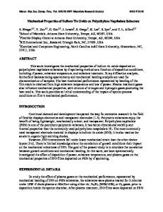
Polyethylene Oxide Nanofibers as Ultraviolet Sensors Saima Naz Khan1, Jeffrey J. Rack2, Aaron R. Rachford2, and Martin E. Kordesch1 1 Physics and Astronomy, Ohio University, 251B Clippinger labs, Athens, OH, 45701 2 Chemistry and Bio-Chemistry, Ohio University, 158 Clippinger labs, Athens, OH, 45701 ABSTRACT Using the electrospinning technique, we have prepared [Ru (pic) 2 (dmso) 2] doped-polyethylene oxide nanofibers for ultraviolet sensing. The diameter of the as-prepared fibers is in the range of 1µm-100nm. These fibers change color from pale yellow to orange when exposed to ultraviolet light (wavelength~350nm) and return to their original color after approximately 2-3 days. The intensity of the color increases with an increased time of exposure to UV. The color changing behavior in the nanofibrous mat is almost the same as that in cast films prepared from the same solution. The scanning electron microscope (SEM) studies of the fibers show that the morphology of the fibers remains unchanged after exposure to UV. INTRODUCTION Electrostatic generation of ultra thin fibers has been known since 1930s [1]. This technique is being used to spin fibers from a wide range of polymer materials [2-3]. Electrospun fibrous mats are used for various applications such as high performance filters [4, 5], and scaffolds in tissue engineering [4, 6] that exploits the high surface area provided by these fibers. One important application of electrospun fibers is made in nanosensors [7] that utilize the electronic properties associated with these fibers. These sensor applications however involve the conductivity measurements before and after the fibers are exposed to the particular environments. In this paper we report for the first time, the electrospinning of nanofibers derived from [Ru (pic) 2 (dmso) 2], a Ruthenium compound that excellent sensors for ultraviolet light (wavelength~350nm). The UV sensing is apparent from the change of color (pale yellow to orange) of these fibers and does not involve any kind of measurements. In the electrostatic technique, a high electric field is generated between the polymer solution contained in a syringe and a grounded metallic collection plate by connecting the needle of the syringe to a high voltage power supply. At a certain critical value of voltage, the polymer droplet is charged enough to overcome the surface tension of the solution and thus a charged jet of the polymer solution is ejected. The jet undergoes a series of electrically induced bending instabilities that result in the stretching of the fibers accompanied by the evaporation of the solvent. The dry fibers are collected on the grounded plate in the form of a non-woven mesh. The schematic of the process is shown in figure 1. EXPERIMENTAL Polyethylene Oxide (PEO) with molecular weight of 300,000 Da was purchased from Aldrich and was used as received to prepare solutions. Chloroform was used as a solvent. [Ru (pic) 2(dmso) 2] powder was obtained from Dr. Jeffrey J. Rack of the Department of Chemistry and
Biochemistry,
Data Loading...