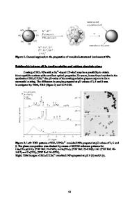
Preparation of CdS/ZnO Core/shell Structured Nanoparticles by Hydrothermal Method
- PDF / 737,460 Bytes
- 5 Pages / 595 x 842 pts (A4) Page_size
- 72 Downloads / 343 Views
Preparation of CdS/ZnO Core/shell Structured Nanoparticles by Hydrothermal Method
Chunhua Yan, Lingdong Sun, Xuefeng Fu, chunsheng Liao State Key Laboratory of Rare Earth Materials Chemistry and Applications, PKU-HKU Joint Laboratory on Rare Earth Materials and Bioinorganic Chemistry, Peking University, Beijing 100871, China
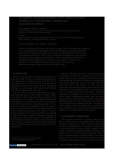
ABSTRACT CdS and ZnO capped CdS (CdS/ZnO) semiconductor nanoparticles were synthesized via hydrothermal method by thermal decomposition of the cysteine-cadmium and Zn(OH)42complex precursors. Both of the photoluminescence properties and structure characterization confirmed the core/shell structure as expected. Compared to CdS nanoparticles, the band-gap emission of CdS/ZnO was greatly improved, that means the capping layer of ZnO modified the surface of CdS and reduced the surface defects effectively. ED and XRD confirmed the formation of hexagon phased CdS and the TEM image indicated the size of CdS/ZnO was about 20 nm. INTRODUCTION Nanosized materials have attracted much consideration in recent years because of the unique properties different very much from the corresponding bulks [1,2]. And nanosized semiconductors have potential applications especially on novel luminescent materials. Many preparation methods have been afforded to improve the quantum efficiency [3-5]. The most successful method is inorganic shell modification, both anionic and cationic sites, are modified by the inorganic shell layer and the surface traps are removed [6-11]. The luminescence efficiency also benefits from the shell and the stability is also increased. We reported on the formation of CdS and CdS/ZnO composite nanoparticles by hydrothermal method, which Zn(OH)42- precursors was used to form ZnO capped CdS nanoparticles. These core/shell nanoparticles were characterized with florescent spectra, transmission electron microscopy (TEM), and X-ray photon spectra (XPS) to study optical properties, morphologies and structures. Compared with CdS nanoparticles, the band-gap emission of CdS/ZnO was greatly improved, that means the capping layer of ZnO modified the surface of CdS and reduced the surface defects effectively. EXPERIMENTAL DETAILS (1) Preparation of CdS nanoparticles 0.6 mL cadmium acetate solution was dropwised into 2.8 mL (0.005 mol/L) L-cysteine solution, and 0.15 mL (0.12 mol/L) NaOH was added to the above mixture to ensure ionization of cysteine to -SCH2CH(NH2)COO-, which can enhance coordination ability to Cd2+. Actually, complex of Cd--SCH2CH(NH2)COO- is used as the precursor to form CdS
H9.41.1
nanoparticles under hydrothermal condition. The reaction temperature and time is controlled to obtain spherical CdS nanoparticles. The resulting yellowish colloid is washed and centrifuged to remove excessive reagents. (2) Preparation of CdS/ZnO nanoparticles 0.4 mL ( 0.050 mol/L) zinc chloride is diluted and pH is set to 12-13 in order to form Zn(OH)42- used as precursors for ZnO capping layer [12]. It is dropwised to the CdSCH2CH(NH2)COO- complex solution used in the above step. The following hydrotherma
Data Loading...